
Dynamic chest radiography could potentially replace pulmonary function tests
New research suggests that dynamic chest radiography is as effective as pulmonary function tests at diagnosing chronic obstructive pulmonary disease.
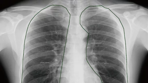
Dynamic chest radiography is a technique that uses continuous radiographic acquisition during respiration to assess lung function. It is easily accessible and when coupled with modern post-processing technology, it can be used to assess changes in the lungs and diaphragm in real-time while patients breathe slowly.
“The technique is easy to operate, requiring only that the patient perform calm breathing and deep breathing maneuvers in front of the detector to obtain image data, and the radiation dose during this examination is comparable to that of conventional frontal and lateral chest radiography or low-dose chest CT examination,” Laiyu Liu, MD, with the department of respiratory and critical care medicine at Nanfang Hospital, Southern Medical University, in China, and colleagues explained. “[Dynamic chest radiography] postprocessing technology can provide precise tracking of diaphragm displacement in the respiratory cycle and can be used to quantitatively assess dynamic changes in lung projection area, providing multidimensional respiratory function parameters for clinical use.”
Typically, pulmonary function tests are used to assess lung function and diagnose COPD. But these exams rely largely on how well patients and providers work together during the test; they also carry a risk of aerosol transmission, which largely hindered their use during the COVID-19 pandemic.
Dynamic chest radiography and pulmonary function tests provide similar diagnostic information on lung function. The emergence of this X-ray method has prompted many to question whether the exam could be used as an alternative to the traditional lung function test when diagnosing COPD. However, research into its utility in this area is limited.
To address this, researchers recently conducted a prospective analysis of 553 individuals with and without COPD between November 2022 and July 2024. Participants underwent both dynamic chest radiography and pulmonary function tests for comparison. Imaging, testing data and patient features were used to make a series of models to predict the likelihood of an individual having COPD.
The team determined that the dynamic chest radiography and pulmonary function tests parameters positively correlated with each other in patients with COPD. The combined bilateral (right and left lungs) rate of change in projected lung area during deep breathing also was linked to forced expiratory volume in 1 second (FEV1) and the ratio of FEV1 to forced vital capacity—an indicator of how well air moves through the lungs. The dynamic chest radiography model, which used three features from imaging, performed well for predicting COPD, but the model that combined imaging parameters with smoking status yielded the most accurate results.
This suggests that dynamic chest radiography "represents an efficacious alternative approach to standard [pulmonary function tests] for COPD screening,” the authors wrote.
“Compared with traditional [pulmonary function tests], [dynamic chest radiography] overcomes the limitations of aerosol transmission and difficult operation, making DCR particularly suitable for the prevention of a high rate of missed diagnosis,” the group noted. “Unlike static imaging, DCR enables real-time visualization of lung and diaphragm kinetics and supports advanced postprocessing to generate functional ventilation and perfusion maps.”
Read more about the findings here.

In addition to her background in journalism, Hannah also has patient-facing experience in clinical settings, having spent more than 12 years working as a registered rad tech. She began covering the medical imaging industry for Innovate Healthcare in 2021.