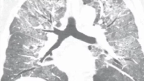
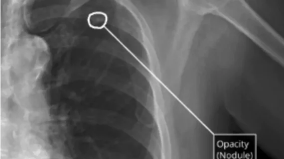
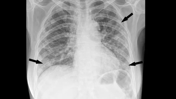
Radiologists use diagnostic imaging to non-invasively look inside the body to help determine the causes of an injury or an illness, and confirm a diagnosis. Providers use many imaging modalities to do so, including CT, MRI, X-ray, Ultrasound, PET and more.
Two months after a cluster of cases occurred on a cruise ship, experts are offering insight into how radiologists can help spot the "silent" signs of the deadly virus on imaging.
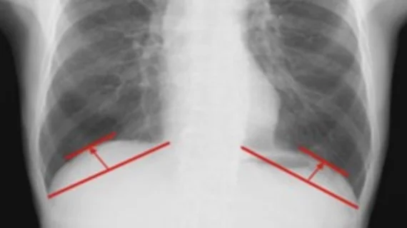
For the first time, research has identified diaphragmatic dome height measured on chest X-rays as an indicator of what patients’ post-op recovery will look like following surgery for cancer or other lung disorders.
On-the-field imaging helps determine if athletes can be treated effectively on-site or whether they might need to be transferred to a medical center for additional care.
A new study has found that a form of liquid biopsy—circulating tumor cells (CTCs)—may be a key technique for creating a staging system to significantly alter the treatment of metastatic breast cancer (MBC).
Currently, unenhanced multi-detector computed tomography (MDCT) is considered the gold standard for detecting kidney stones, however the modality also delivers the highest radiation dose among imaging methods.
A group of German researchers found diffusion-weighted (DW) MRI provided superior prognostic information compared to PET/CT in liver cancer patients who underwent 90Y radioembolization and proved more accurate in predicting overall survival in these patients.
Using functional MRI (fMRI), researchers from VU University Medical Center in Amsterdam found a correlation between white matter brain damage and atrophy in multiple sclerosis (MS) patients—a primary factor of cognitive impairment in patients with the disease.
New research has found fetal MRI can reliably identify holoprosencephaly as early as 18 weeks into pregnancy, providing added time for parents to understand and prepare for the condition.
Microsoft will collaborate with Case Western Reserve University in Cleveland to improve the accuracy of MRI results in less time through an approach called "magnetic resonance fingerprinting."
NASA astronauts at the International Space Station used portable ultrasound to scan each other for spinal cord changes that may occur during long-term space missions, according to a study published in the Journal of Ultrasound in Medicine.
Researchers at CalTech are developing "erasable" contrast agents that can "blink off" on command during an MRI to reveal their exact location inside the body, according to a press release from the California Institute of Technology.