
Hantavirus on imaging: What radiologists need to know
Two months after news broke of a hantavirus subtype spreading among cruise ship passengers, experts are offering insight into how radiologists can help spot the signs of the deadly virus on imaging.
In early April, reports pertaining to the cruise ship MV Hondius, which set sail out of Argentina, began to circulate after several patients started to exhibit concerning symptoms. One 70-year-old man experienced severe respiratory and gastrointestinal issues, which eventually progressed to respiratory distress. He eventually died on the ship five days after his symptoms first emerged. The septuagenarian is one of three passengers, including his wife, who passed away after contracting what health authorities determined was a hantavirus subtype capable of spreading between humans. Dozens of others fell ill and were forced to quarantine.
Hantavirus, specifically the Andes virus (ANDV), is rare but deadly. It typically is categorized into two overlapping syndromic patterns: hemorrhagic fever with renal syndrome (HFRS) and hantavirus pulmonary syndrome (HPS), also termed hantavirus cardiopulmonary syndrome (HCPS). It causes flu-like symptoms that can rapidly progress to the point of respiratory distress and kidney failure. For this reason, identifying individuals who have been infected early on is critical to prevent further spread. Rads play a key role in this, authors of a new paper in Emergency Radiology contend.
“Radiologists, particularly those in emergency and thoracic imaging, are often the first to identify the ‘silent’ imaging patterns suggestive of hantavirus infection,” Ali Gholamrezanezhad, with the department of radiology at Keck Medicine of USC, Los Angeles, and colleagues noted. “Consequently, a detailed understanding of the imaging manifestations, differential diagnoses, and stringent infection-control considerations is essential for the modern emergency radiology practice.”
The research team reviewed all available data on confirmed hantavirus cases, pinpointing those with imaging exams to review. Using this information, they compiled a list of imaging manifestations unique to the virus that radiologists can use as a reference when interpreting exams from patients exposed to the virus.
Here’s what they came up with:
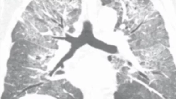
Standard chest X-rays are the preferred first-line imaging modality for hantavirus. CT is not required for diagnosing the virus but can be useful when the decision is in question or when symptoms have progressed.
The hallmark finding of hantavirus pulmonary syndrome is non-cardiogenic pulmonary edema. Bilateral ground-glass opacities, smooth interlobular and intralobular septal thickening and peribronchovascular thickening are common in cases of the syndrome.
Pleural effusions are commonly found in severe hantavirus pulmonary syndrome cases.
These findings, alongside a normal heart size, are especially indicative of the syndrome.
For hemorrhagic fever with renal syndrome cases, renal enlargement and retroperitoneal edema are common. Hemorrhagic complications have been documented in severe cases.
The team acknowledged that many of these findings overlap with other clinical causes. As such, they should be interpreted alongside a thorough clinical history that includes blood work, information on recent travel and any knowledge of potential exposure to others who may have come in contact with the virus.
“For emergency medicine and radiology teams, [hantavirus pulmonary syndrome] moves quickly and brings tough challenges,” the authors warned. “Early recognition of characteristic imaging findings can help doctors get patients to the ICU and start advanced care before their lungs fail. Late diagnosis can overwhelm emergency and critical care services especially in places with limited resources.”
Read more here.

In addition to her background in journalism, Hannah also has patient-facing experience in clinical settings, having spent more than 12 years working as a registered rad tech. She began covering the medical imaging industry for Innovate Healthcare in 2021.