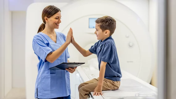
Radiologists use diagnostic imaging to non-invasively look inside the body to help determine the causes of an injury or an illness, and confirm a diagnosis. Providers use many imaging modalities to do so, including CT, MRI, X-ray, Ultrasound, PET and more.
Chest CT is routinely performed for things like lung cancer screening, coronary calcium assessment and pulmonary nodule surveillance, but the modality also can be used to extract additional information.
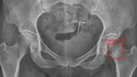
By highlighting subtle abnormalities that could otherwise be overlooked, an algorithm could potentially serve as an additional safety net when radiographic findings are indeterminate.
Despite clinical guidelines, the use of advanced imaging for patients complaining of headaches has increased, according to a study published online Jan. 8 in the Journal of General Internal Medicine.
The latest version of Siemens Healthcare’s Multix Fusion digital radiography system has received 510(k) clearance from the FDA and is now available in the U.S.
Time magazine compiled its list of last year’s remarkable healthcare innovations and achievements, and No. 1 was 3D mammography, or tomosynthesis.
Race and ethnicity play a role in both early diagnosis of invasive breast cancer and survival rates after diagnosis, according to a study published online Jan. 13 in the Journal of the American Medical Association.
A follow-up chest CT after abnormal x-ray findings produces a high yield of relevant findings, according to a study published online in Radiology on Dec. 22.