Radiologists use diagnostic imaging to non-invasively look inside the body to help determine the causes of an injury or an illness, and confirm a diagnosis. Providers use many imaging modalities to do so, including CT, MRI, X-ray, Ultrasound, PET and more.
Chest CT is routinely performed for things like lung cancer screening, coronary calcium assessment and pulmonary nodule surveillance, but the modality also can be used to extract additional information.
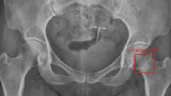
By highlighting subtle abnormalities that could otherwise be overlooked, an algorithm could potentially serve as an additional safety net when radiographic findings are indeterminate.
Communication is the core reasoning behind setting up a 3D lab, according to Matthew A. Barish, MD, of Brigham and Women's Hospital who presented Setting up a 3D lab on Friday.
FUJIFILM Medical Systems USA, Inc. said Friday it has submitted the final module of its pre-market approval (PMA) application with the FDA for the approval of Fuji Computed Radiography for Mammography (FCRm).
Siemens is touting the benefits of its SIENET Cosmos web-based RIS-PACS with integrated digital dictation and voice functions - which will be installed in 40 facilities by year's end.