Radiologists use diagnostic imaging to non-invasively look inside the body to help determine the causes of an injury or an illness, and confirm a diagnosis. Providers use many imaging modalities to do so, including CT, MRI, X-ray, Ultrasound, PET and more.
Chest CT is routinely performed for things like lung cancer screening, coronary calcium assessment and pulmonary nodule surveillance, but the modality also can be used to extract additional information.
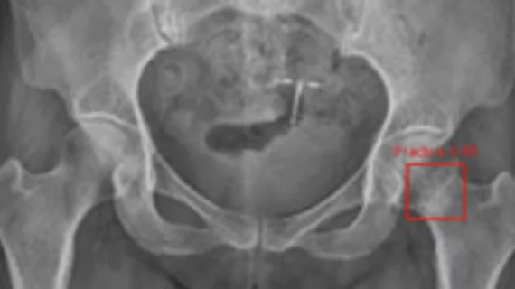
By highlighting subtle abnormalities that could otherwise be overlooked, an algorithm could potentially serve as an additional safety net when radiographic findings are indeterminate.
Cedara Software Corp. on Tuesday signed a $48 million agreement to acquire RIS, PACS and teleradiology provider, eMed Technologies Corp. of Burlington, Mass.
Women with a high genetic risk for breast cancer run a better chance of having it detected with MRI than with mammography and other methods, according to a study published in this week's Journal of the American Medical Association.
Reid Hospital and Healthcare Services in Richmond, Ind., is the first hospital to deploy and utilize Toshiba America Medical Systems Inc.'s (TAMS) new cardiac flat-panel detector (FPD) for the Infinix-i cardiovascular X-ray system.
Shands Hospital at the University of Florida (Shands at UF), a teaching hospital of the Shands HealthCare system, has selected HIPAAT Inc.'s products to assist with HIPAA security and privacy efforts.
The Angiography and X-ray Division of Siemens Medical Solutions has introduced interventional cardiac 3D (IC3D) technology to create three-dimensional (3D) images of the coronary vessels in the heart.