Radiologists use diagnostic imaging to non-invasively look inside the body to help determine the causes of an injury or an illness, and confirm a diagnosis. Providers use many imaging modalities to do so, including CT, MRI, X-ray, Ultrasound, PET and more.
Chest CT is routinely performed for things like lung cancer screening, coronary calcium assessment and pulmonary nodule surveillance, but the modality also can be used to extract additional information.
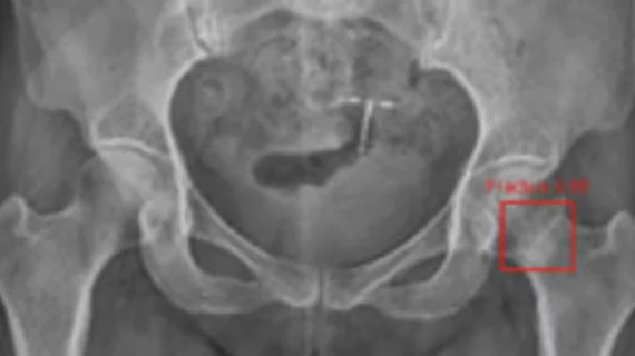
By highlighting subtle abnormalities that could otherwise be overlooked, an algorithm could potentially serve as an additional safety net when radiographic findings are indeterminate.
Varian Medical Systems, Inc. has developed a new metal technology replacement tube for use with cardiac catheterization lab equipment from Siemens Medical Solutions.
Surgical teams at Henry Ford Hospital's Vattikuti Urology Institute are now performing robot-assisted prostatectomy using revolutionary three-dimensional (3D) technology in a newly constructed operating room (OR).
TeraRecon, Inc. has entered the flat-panel detector market after this week's announcement of a strategic investment in Acrorad Co. Ltd., Okinawa, Japan.