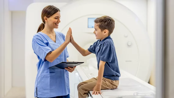
Radiologists use diagnostic imaging to non-invasively look inside the body to help determine the causes of an injury or an illness, and confirm a diagnosis. Providers use many imaging modalities to do so, including CT, MRI, X-ray, Ultrasound, PET and more.
Chest CT is routinely performed for things like lung cancer screening, coronary calcium assessment and pulmonary nodule surveillance, but the modality also can be used to extract additional information.
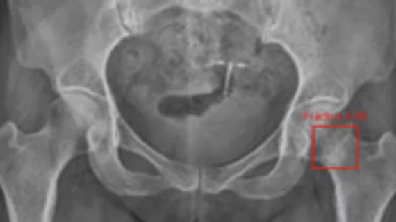
By highlighting subtle abnormalities that could otherwise be overlooked, an algorithm could potentially serve as an additional safety net when radiographic findings are indeterminate.
Increasing knowledge of the human genome, wider acceptance of digital and wireless technology, and the use of advanced imaging systems are rapidly changing the face of the healthcare industry.
In collaboration with the International Task Force for Prevention of Coronary Heart Disease, Siemens Medical Solutions introduced a refined method for the assessment of a patient's risk for coronary heart disease (CHD).
Bristol-Myers Squibb's (BMS) Medical Imaging division released new findings at ACC 2004 that suggest more than one-third of patients hospitalized at the initial onset of heart failure who undergo myocardial perfusion imaging (MPI) with Cardiolite may not
Revenues and earnings at Analogic Corp. declined in its second fiscal quarter, ending Jan. 31, as growth in medical imaging equipment could not offset a drop in explosive detection technology.