Magnetic Resonance Imaging

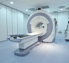
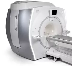
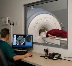
Magnetic resonance imaging (MRI) is the gold standard imaging modality for soft tissues. It produces detail cross-sectional images of soft tissue and bone anatomy, including muscles, tendons, ligaments, brain and organs, without the use of ionizing radiation. In addition to orthopedic imaging, MRI is also used for heart, brain and breast. MRI uses gadolinium contrast in many exams to highlight tissues and blood vessels, which enhances images and offers better diagnostic quality. It can also be used in conjunction with PET scans. How does MRI work? MR creates images by using powerful magnets to polarize hydrogen atoms in water (the body is made of of more than 80% water) so they face in one direction. A radiofrequency pulse is then used to ping these atoms, causing them to wobble, or resonate. The MRI coils detect this and computers can assemble images from the signals. Basic MRI scans will focus on the resonance of fat and water in two different sequences, which highlight and contrast different features in the anatomy.

Thumbnail

With safety events on the rise, new board will credential for MR safety

An organization called the American Board of Magnetic Resonance Safety (ABMRS) has been formed with a mission to certify—through formally administered tests—individuals who have been delegated the responsibility of ensuring the safety of clinical and research magnetic resonance environments.

Thumbnail

New York radiology practice replaces two MR systems with one; Significantly grows volumes

Sponsored by Hitachi Healthcare Americas

Serving the community of upstate New York’s Capital Region, Adirondack Radiology Associates (ARA) recently expanded patients’ access to state-of-the-art, high-field open bore MR imaging technology, replacing two systems, an older low-field open system and a closed 1.5T system, with the Hitachi Oasis high-field open MR at their Saratoga Imaging Center. Since the installation, ARA has experienced an increase in procedure volumes and patient satisfaction as well a reduction in costs. 

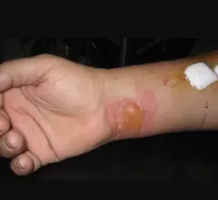
Example of a RF burn from an MRI scan where a sedated patient's identification bracelet was touching their skin during an exam. Image courtesy of RSNA. https://pubs.rsna.org/doi/10.1148/radiol.09090637

Battling Burns in MRI

While MRI is often considered one of the safer imaging modalities due to its lack of ionizing radiation, other dangers remain for patients, with thermal burns being a chief concern. 

Thumbnail

GE Healthcare’s New SIGNA™ PET/MR Pending 510(k) Approval

Waukesha, WI — August 4, 2014 — Today GE Healthcare announced that the first integrated, simultaneous, time-of-flight (TOF) capable, whole body SIGNA PET/MR* is 510(k) pending at the FDA. Powered by simultaneous image acquisition from GE’s latest 3.0 Tesla magnetic resonance (MR) technology and innovative positron emission tomography (PET) technology, the SIGNA PET/MR represents a new chapter in helping clinicians achieve improved scan efficiency that may lead to more effective treatment paths for clinicians to offer their patients, particularly for oncology, neurology, and cardiology.

Thumbnail

Steering patients to low-cost facilities lowered MRI costs by 18.7 percent

A radiology benefits management (RBM) company reduced the average cost of an MRI by 9 percent in markets where patients received phone calls directing them to lower-cost facilities, according to a study published in Health Affairs.

Thumbnail

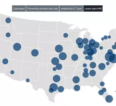
Cost comparison map underscores price variation for MRI, CT

Sacramento leads the country in pricing for an MRI of the lower back (average $2,365), and just a short jaunt up the West Coast, Seattle offers the nation’s lowest average cost for a back scan at $907, according to a new analysis of in-network price differences for four common medical procedures.

Thumbnail

G.E. Healthcare partners with Tesla on 7T MRI research scanners

G.E. Healthcare, Buckinghamshire, U.K., announced a partnership with West Sussex, U.K.-based Tesla Engineering Ltd. on the development of a new 7T whole-body MRI scanner.

Thumbnail

Increased Access, Dependability and Patient Satisfaction with a Fixed MRI at Star Valley CAH

Sponsored by Hitachi Healthcare Americas

Among the many requirements a hospital must meet to achieve and maintain the designation of Critical Access Hospital (CAH), the hospital must be fairly remote and furnish 24-hour emergency care services seven days a week, using either on-site or on-call staff.

Around the web

The nuclear imaging isotope shortage of molybdenum-99 may be over now that the sidelined reactor is restarting. ASNC's president says PET and new SPECT technologies helped cardiac imaging labs better weather the storm.

CMS has more than doubled the CCTA payment rate from $175 to $357.13. The move, expected to have a significant impact on the utilization of cardiac CT, received immediate praise from imaging specialists.

The all-in-one Omni Legend PET/CT scanner is now being manufactured in a new production facility in Waukesha, Wisconsin.