Magnetic Resonance Imaging

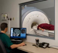
Magnetic resonance imaging (MRI) is the gold standard imaging modality for soft tissues. It produces detail cross-sectional images of soft tissue and bone anatomy, including muscles, tendons, ligaments, brain and organs, without the use of ionizing radiation. In addition to orthopedic imaging, MRI is also used for heart, brain and breast. MRI uses gadolinium contrast in many exams to highlight tissues and blood vessels, which enhances images and offers better diagnostic quality. It can also be used in conjunction with PET scans. How does MRI work? MR creates images by using powerful magnets to polarize hydrogen atoms in water (the body is made of of more than 80% water) so they face in one direction. A radiofrequency pulse is then used to ping these atoms, causing them to wobble, or resonate. The MRI coils detect this and computers can assemble images from the signals. Basic MRI scans will focus on the resonance of fat and water in two different sequences, which highlight and contrast different features in the anatomy.

Caught in the act: Man sentenced for acquiring Oxycodone with fake MRI results

James Lawlor was sentenced to a year of supervised release this week for his role in a large-scale scam that involved using fake MRI results to acquire Oxycodone. This comes months after other participants were sentenced for their own involvement.

UT Dallas professor earns prestigious award from ISMRM

A. Dean Sherry has been awarded the Gold Medal Award by the International Society for Magnetic Resonance in Medicine (ISMRM) for significant achievements made over the course of his 40-year career. 

Nanotechnology identifies brain tumor types through MRI 'virtual biopsy'

LOS ANGELES (May 26, 2015) – Biomedical researchers at Cedars-Sinai have invented a tiny drug-delivery system that can identify cancer cell types in the brain through “virtual biopsies” and then attack the molecular structure of the disease.

New DTN protocol renders MRI effective for screening stroke patients

Thanks to reduced door-to-needle (DTN) time, MRI was proven to be an effective screening tool for stroke victims before they receive acute treatment, according to findings published in Neurology. 

Thumbnail

Patient motion during MRI is $115,000 problem

Patient motion during MRI is costing healthcare providers an estimated $115,000 per scanner per year, according to a study published in the Journal of the American College of Radiology.

Thumbnail

Bill would OK privately owned MRI scanners in Saskatchewan—with a catch

Saskatchewan Health Minister Dustin Duncan has introduced a new bill that would make Saskatchewan the latest Canadian province to allow privately owned MRIs. 

Thumbnail

High Precision Devices receives Excellence in Technology Transfer Award for quantitative MRI phantom

Boulder, Colorado May 6, 2015 /PRNewswire/ High Precision Devices, Inc. (HPD) received a joint award with the National Institute of Standards and Technology (NIST) for commercializing the temperature-controlled spherical magnetic resonance imaging phantom, branded as the HPD Diffusion Phantom Model 128.

Thumbnail

Vermont tests elimination of payer pre-approvals for MRIs

The Green Mountain State is in the news for more than the entry of Independent Sen. Bernie Sanders into the 2016 race for president. A pilot healthcare-delivery project there experiments with removing a rule currently requiring patients to get pre-approval from their health plans for MRIs. 

Around the web

The nuclear imaging isotope shortage of molybdenum-99 may be over now that the sidelined reactor is restarting. ASNC's president says PET and new SPECT technologies helped cardiac imaging labs better weather the storm.

CMS has more than doubled the CCTA payment rate from $175 to $357.13. The move, expected to have a significant impact on the utilization of cardiac CT, received immediate praise from imaging specialists.

The all-in-one Omni Legend PET/CT scanner is now being manufactured in a new production facility in Waukesha, Wisconsin.