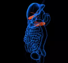
Magnetic Resonance Imaging

Magnetic resonance imaging (MRI) is the gold standard imaging modality for soft tissues. It produces detail cross-sectional images of soft tissue and bone anatomy, including muscles, tendons, ligaments, brain and organs, without the use of ionizing radiation. In addition to orthopedic imaging, MRI is also used for heart, brain and breast. MRI uses gadolinium contrast in many exams to highlight tissues and blood vessels, which enhances images and offers better diagnostic quality. It can also be used in conjunction with PET scans. How does MRI work? MR creates images by using powerful magnets to polarize hydrogen atoms in water (the body is made of of more than 80% water) so they face in one direction. A radiofrequency pulse is then used to ping these atoms, causing them to wobble, or resonate. The MRI coils detect this and computers can assemble images from the signals. Basic MRI scans will focus on the resonance of fat and water in two different sequences, which highlight and contrast different features in the anatomy.

Thumbnail

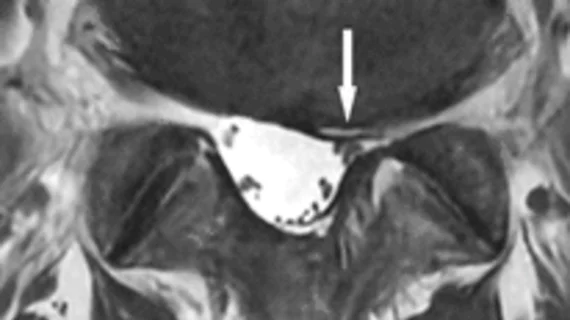
Vermont pilot program averts pre-auth for select spine MRIs

Two medical centers in Vermont kick off a new pilot program on May 1 that circumvents the onerous pre-authorization process by preapproving selected lumbar spine MRIs.

Thumbnail

MRI-only screening of those at risk of pancreatic cancer shows promise

A new study from Karolinska Institute in Stockholm, Sweden, demonstrated that a noninvasive screening protocol of MRI alone in individuals at high risk of pancreatic cancer resulted in a lesion yield of 40% in the screened patients.

FDA recalls GE, Siemens MRI systems

The Food and Drug Administration has issued two separate Class 2 recalls involving Siemens and GE MRI systems.

Thumbnail

Got an EMR? Get an enterprise-wide ‘clinical imaging health record’

At a time when 85% of physicians are viewing diagnostic images via electronic medical records, the opportunity is ripe to realize true enterprise imaging. More of those docs should be saying goodbye to walled-off image silos and hello to a centralized, always-open image depot. 

MRI technique distinguishes cancerous from noncancerous cells

A new magnetic resonance imaging technique could potentially make biopsies obsolete by noninvasively detecting sugar molecules that are shed by the outer membranes of cancerous cells.

Thumbnail

We’re all MRU experts now

The medical imaging community is buzzing over the GE MRI recall—by now, no other words are needed to name it—and the chatter will probably continue for a long time to come.

GE recalls thousands of MRIs

GE is recalling almost 13,000 MRI units, as the FDA has deemed them potentially deadly.

FDA recall notice follows GE letter advising customers to check MRI systems

Citing issues with how the magnetic rundown unit (MRU) is connected to the magnet at some MRI sites, the Food and Drug Administration (FDA) has issued a Class 1 recall for all GE Healthcare MRIs with superconducting magnets, and GE Healthcare is asking its customers to perform a test to ensure that the MRU is properly connected.

Around the web

The nuclear imaging isotope shortage of molybdenum-99 may be over now that the sidelined reactor is restarting. ASNC's president says PET and new SPECT technologies helped cardiac imaging labs better weather the storm.

CMS has more than doubled the CCTA payment rate from $175 to $357.13. The move, expected to have a significant impact on the utilization of cardiac CT, received immediate praise from imaging specialists.

The all-in-one Omni Legend PET/CT scanner is now being manufactured in a new production facility in Waukesha, Wisconsin.