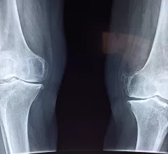
Orthopedic imaging relies on X-ray, MRI and CT to diagnose disorders and injuries affecting the bones, muscles, ligaments, tendons, cartilage, and spine. Orthopedists also use these test results to create an effective treatment plan.
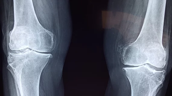
Low-dose radiation therapy may do more than relieve pain in patients with early knee osteoarthritis, with new long-term findings suggesting the treatment could also slow structural deterioration.
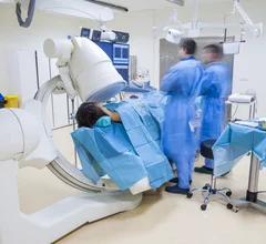
The procedure reduces inflammation and pain, and allows patients to achieve relief without having to undergo surgery and months of physical therapy during recovery.
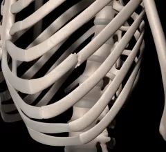
“These findings emphasize the importance of early recognition of IPV and timely intervention to prevent further harm to the victim,” authors of research published in Academic Radiology cautioned.
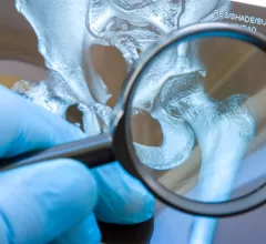
Because sacral insufficiency fractures do not always show a fracture line, they can be difficult to diagnose or even misdiagnosed as bone metastasis, which could result in additional treatments like radio-chemotherapy.
A study in Lancet Digital Health reports that a previously validated, high performing AI model committed troublesome errors when confronted with atypical anatomy.
Experts urged physicians to take extra protective measures when in the presence of metal protheses during procedures and to be vigilant in shielding their eyes from additional exposure.
Research published recently in Radiology found comparable sensitivity and specificity between artificial intelligence and clinicians for fracture detection.
Many decision support tools catered to knee osteoarthritis have emerged in recent years, but external validation that ensures these algorithms can operate in a clinical setting has been lacking.