Breast Imaging

Breast imaging includes imaging modalities used for breast cancer screenings and planning therapy once cancer is detected. Mammography is the primary modality used. Mammogram technology is moving from 2D full-field digital mammography (FFDM) to breast tomosynthesis, or 3D mammography, which helps reduce false positive exams by allowing radiologists to look through the layers of tissue. Overlapping areas of dense breast tissue on 2D mammograms appear similar to cancers and 3D tomo helps determine if suspect areas are cancer or not. About 50% of women have dense breast tissue, which appears white on mammograms, the same as cancers, making diagnosis difficult. Radiologists use the Breast Imaging Reporting and Data System (BI-RADS) scoring system to define the density of breast tissue. Many states now require patients to be notified if they have dense breasts so they understand their mammograms might be suboptimal and they should use supplemental imaging that can see through the dense areas. This includes tomosythesis, breast ultrasound, automated breast ultrasound (ABUS), breast MRI, contrast enhanced mammography and nuclear imaging, including positron emission mammography (PEM).
Breast cancer AI ribbon pink artificial intelligence

AI effectively flags mammograms of women who would benefit from supplemental MRI

Experts involved in the algorithm's development believe its time-saving potential could help improve both radiologist workflows and patient outcomes. 

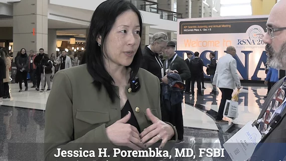
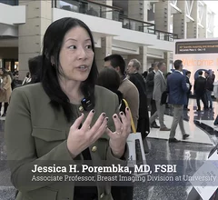
Jessica H. Porembka, MD, FSBI, associate professor, breast imaging division University of Texas Southwestern Medical Center, Dallas, and vice chair of strategy and quality, and quality assurance medical director, Parkland Radiology in Dallas, explains how an ultrasound-first strategy for noncalcified lesions in DBT proves cost-effective.

Ultrasound-first strategy for noncalcified lesions in DBT proves cost-effective

Jessica Porembka, MD, of the breast imaging division at University of Texas Southwestern Medical Center, said an ultrasound-first strategy for these lesions in DBT is cost-effective and improves efficiency. 

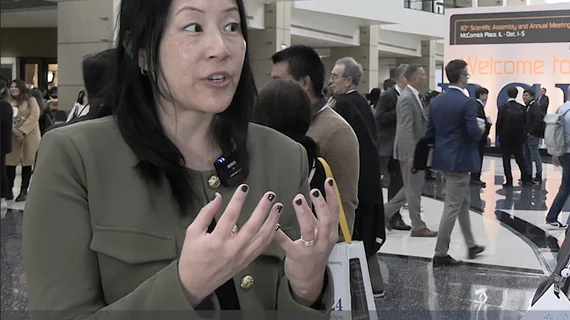
Jessica H. Porembka, MD, FSBI, associate professor, breast imaging division University of Texas Southwestern Medical Center, Dallas, and vice chair of strategy and quality, and quality assurance medical director, Parkland Radiology in Dallas, discusses what it means when a radiology report says a patient has an architectural distortion on their mammogram.

What patients need to known about architectural distortion on breast imaging

Radiologist Jessica Porembka, MD, FSBI, an associate professor with the breast imaging division at University of Texas Southwestern Medical Center, explains what it means when a mammography report says a patient has architectural distortion.
 

PHOTO GALLERY: Medical imaging technology at RSNA 2024

Take a virtual tour around the Radiological Society of North America (RSNA) meeting to see the sights and new technologies displayed across the vast exhibit hall floors.

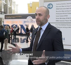
 RBMA Board member Kit Crancer outlines the key legislation in radiology to watch.

Pending radiology bills in Congress and predicting the Trump administration's impact on imaging

Kit Crancer, RBMA board member, speaks with Radiology Business about key legislative developments on the Hill that will affect the specialty. 

breast cancer screening mammography

AI accurately predicts breast cancer years before diagnosis

This information could help providers personalize breast cancer screening strategies and initiate treatment earlier.

Thumbnail

Radiology AI firm specializing in automating MRI workflows raises $19M

Copenhagen-based Cerebriu said the Danish VC firm North Ventures led the Series A funding round with support from EIFO and Sagitta Ventures. 

Video interview with Debra Monticciolo, MD, on the expansion of DBT and reducing breast imaging recall rates.

Breast tomosynthesis reduces recalls, could soon replace routine 2D mammography

Digital breast tomosythesis now makes up close to 50% of mammography systems in the U.S. "Tomo is going to replace just straight digital mammography simply because of the benefit of fewer recalls," explained Debra L. Monticciolo, MD.

Around the web

The patient, who was being cared for in the ICU, was not accompanied or monitored by nursing staff during his exam, despite being sedated.

The nuclear imaging isotope shortage of molybdenum-99 may be over now that the sidelined reactor is restarting. ASNC's president says PET and new SPECT technologies helped cardiac imaging labs better weather the storm.

CMS has more than doubled the CCTA payment rate from $175 to $357.13. The move, expected to have a significant impact on the utilization of cardiac CT, received immediate praise from imaging specialists.