Breast Imaging

Breast imaging includes imaging modalities used for breast cancer screenings and planning therapy once cancer is detected. Mammography is the primary modality used. Mammogram technology is moving from 2D full-field digital mammography (FFDM) to breast tomosynthesis, or 3D mammography, which helps reduce false positive exams by allowing radiologists to look through the layers of tissue. Overlapping areas of dense breast tissue on 2D mammograms appear similar to cancers and 3D tomo helps determine if suspect areas are cancer or not. About 50% of women have dense breast tissue, which appears white on mammograms, the same as cancers, making diagnosis difficult. Radiologists use the Breast Imaging Reporting and Data System (BI-RADS) scoring system to define the density of breast tissue. Many states now require patients to be notified if they have dense breasts so they understand their mammograms might be suboptimal and they should use supplemental imaging that can see through the dense areas. This includes tomosythesis, breast ultrasound, automated breast ultrasound (ABUS), breast MRI, contrast enhanced mammography and nuclear imaging, including positron emission mammography (PEM).
breast ultrasound biopsy

Reducing breast cancer care costs via triage with AI-ultrasound combo

Artificial intelligence applied to portable breast US can reduce about half of unnecessary referrals for benign lesions, experts wrote in Radiology

breast radiologist breast cancer mammography

Providers chart uptick in number of breast MRI denials issued by insurers

Among vulnerable women with BRCA1/2 mutations, about 14% never received this vital exam after the rejection, MSK researchers reported.

radiology reporting EHR health record CDS AUC

Lessons learned from 7 years of structured radiology reporting at 1 institution

The University Medical Center Mainz recently surveyed radiologists and referrers to gather feedback on the change. 

mammography mammogram breast cancer

Nearly 100,000 individuals sign petition demanding alternative to painful mammography

Dutch woman Murial van der Draaij first launched the effort, characterizing the traditional compression exam as "torture." 

breast ultrasound biopsy

‘Startling’ study findings show persistent barrier to follow-up imaging after a mammogram

Women covered by commercial plans with higher out-of-pocket costs received significantly fewer subsequent procedures, experts wrote Monday in JAMA Network Open.  

Telemammography

4 key trends in breast imaging

These trends include growth in 3D mammography, supplemental imaging for women with dense breasts and in the role of artificial intelligence.

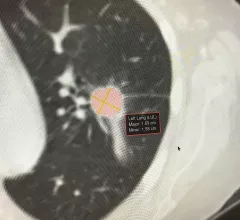
Example of artificial intelligence generated measurements to quantify the size of a lung cancer nodule during a followup CT scan to see if the lesion is regressing with treatment. This type of automation can aid radiologists by doing the tedious, time consuming work. Photo by Dave Fornell

8 trends in radiology technology to watch in 2023

Here is a list of some key trends in radiology technology from our editors based on our coverage of the radiology market.

 Winthrop P. Rockefeller Cancer Institute, University of Arkansas for Medical Sciences (USMS), is addressing health inequities in mammography using its Mammovan mobile breast imaging screening program. Gwendolyn Bryant-Smith, MD, explained how the program works. #RSNA #RSNA22

VIDEO: Bringing health equity to mammography and health screenings in Arkansas

Gwendolyn Bryant-Smith, MD, division chief of breast imaging and associate director for diversity, equity and inclusion at the Winthrop P. Rockefeller Cancer Institute, University of Arkansas, discussed how her center addressed health inequities in mammography.

Around the web

The ACR hopes these changes, including the addition of diagnostic performance feedback, will help reduce the number of patients with incidental nodules lost to follow-up each year.

And it can do so with almost 100% accuracy as a first reader, according to a new large-scale analysis.

The patient, who was being cared for in the ICU, was not accompanied or monitored by nursing staff during his exam, despite being sedated.