Breast Imaging

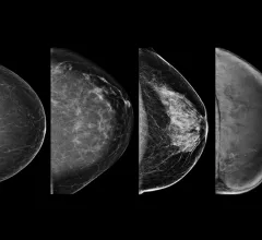
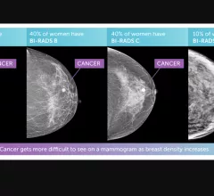
Breast imaging includes imaging modalities used for breast cancer screenings and planning therapy once cancer is detected. Mammography is the primary modality used. Mammogram technology is moving from 2D full-field digital mammography (FFDM) to breast tomosynthesis, or 3D mammography, which helps reduce false positive exams by allowing radiologists to look through the layers of tissue. Overlapping areas of dense breast tissue on 2D mammograms appear similar to cancers and 3D tomo helps determine if suspect areas are cancer or not. About 50% of women have dense breast tissue, which appears white on mammograms, the same as cancers, making diagnosis difficult. Radiologists use the Breast Imaging Reporting and Data System (BI-RADS) scoring system to define the density of breast tissue. Many states now require patients to be notified if they have dense breasts so they understand their mammograms might be suboptimal and they should use supplemental imaging that can see through the dense areas. This includes tomosythesis, breast ultrasound, automated breast ultrasound (ABUS), breast MRI, contrast enhanced mammography and nuclear imaging, including positron emission mammography (PEM).
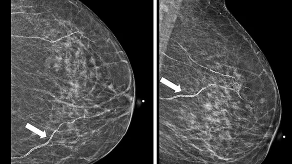
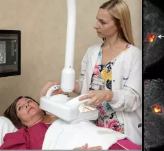
Supplemental breast imaging modalities of automated breast ultrasound (ABUS) and molecular imaging.

VIDEO: Use of supplemental imaging in women with dense breasts

Society of Breast Imaging (SBI) President John Lewin, MD, discusses the types of supplemental breast imaging used to aid cancer detection in women with dense breast tissue.

Example of an artificial intelligence (AI) app store on the Sectra website, where Sectra PACS users can select the AI algorithms they want that are already integrated into the Sectra System. Other vendors have followed a similar approach to AI developed by many smaller vendors they partner with.

VIDEO: Development of AI app stores to enable easier access

Keith J. Dreyer, DO, PhD, FACR, American College of Radiology (ACR) Data Science Institute Chief Science Officer, explains how radiology vendors have developed AI app stores to make it easier to access new FDA cleared AI algorithms.
 

Keith J. Dreyer, DO, PhD, FACR, American College of Radiology (ACR) Data Science Institute Chief Science Officer, explains artificial intelligence (AI) for radiology. Dreyer also holds the positions of vice chairman of radiology at Massachusetts General Hospital, chief data science and information officer for the departments of radiology for both Massachusetts General Hospital and Brigham and Women's Hospital.

VIDEO: Where will radiology AI be in 5 years?

Keith J. Dreyer, DO, PhD, FACR, American College of Radiology (ACR) Data Science Institute Chief Science Officer, explains 5 developments to watch for in radiology artificial intelligence (AI).

COVID-19 medical imaging examples of various clinical presentations. SARS-CoV-2 clinical imaging presentations.

PHOTO GALLERY: What does COVID-19 look like on medical imaging?

This image gallery shows what the various clinical presentations associated with the COVID-19 virus that have been documented during the coronavirus pandemic.

Thumbnail

Deep learning expedites normal findings on ultrafast breast screenings

AI can safely and accurately identify healthy breast tissue on ultrafast breast MRI, negating the need for a radiologist’s closer look and, in the process, lowering cancer screening costs and widening patient access to breast MRI.

breast cancer screening mammography

Malignant architectural distortion ably diagnosed on breast imaging by human-AI combo

Combining ensemble AI models with reads from breast radiologists of mixed experience levels can help health systems consistently diagnose malignant architectural distortion on mammography.

Thumbnail

Abbreviated breast MRI deemed an attractive screening option—sometimes

AB-MRI is a cost-effective means of screening women with dense breast tissue for breast cancer—as long as the per-exam costs don’t top 82% of what would have been spent to perform full-protocol breast MRI.

breast radiologist breast cancer mammography

Screening breast MRI results in more downstream healthcare costs than mammography alone

Women who underwent magnetic resonance imaging experienced more subsequent scans, procedures, healthcare visits and hospitalizations. 

Around the web

The nuclear imaging isotope shortage of molybdenum-99 may be over now that the sidelined reactor is restarting. ASNC's president says PET and new SPECT technologies helped cardiac imaging labs better weather the storm.

CMS has more than doubled the CCTA payment rate from $175 to $357.13. The move, expected to have a significant impact on the utilization of cardiac CT, received immediate praise from imaging specialists.

The all-in-one Omni Legend PET/CT scanner is now being manufactured in a new production facility in Waukesha, Wisconsin.

Trimed Popup
Trimed Popup