Breast Imaging

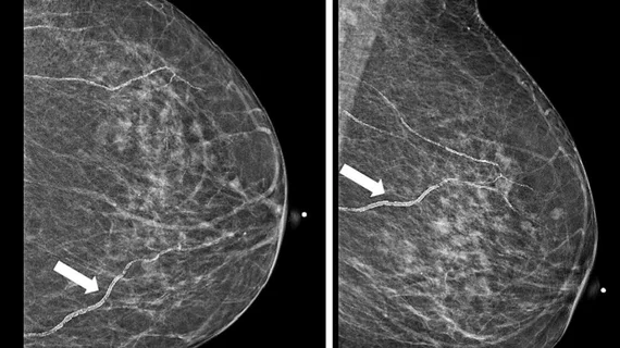
Breast imaging includes imaging modalities used for breast cancer screenings and planning therapy once cancer is detected. Mammography is the primary modality used. Mammogram technology is moving from 2D full-field digital mammography (FFDM) to breast tomosynthesis, or 3D mammography, which helps reduce false positive exams by allowing radiologists to look through the layers of tissue. Overlapping areas of dense breast tissue on 2D mammograms appear similar to cancers and 3D tomo helps determine if suspect areas are cancer or not. About 50% of women have dense breast tissue, which appears white on mammograms, the same as cancers, making diagnosis difficult. Radiologists use the Breast Imaging Reporting and Data System (BI-RADS) scoring system to define the density of breast tissue. Many states now require patients to be notified if they have dense breasts so they understand their mammograms might be suboptimal and they should use supplemental imaging that can see through the dense areas. This includes tomosythesis, breast ultrasound, automated breast ultrasound (ABUS), breast MRI, contrast enhanced mammography and nuclear imaging, including positron emission mammography (PEM).
breast ultrasound biopsy

3 more states pass legislation bolstering breast imaging coverage

New Hampshire is one of the latest after Republican Gov. Chris Sununu signed House Bill 1296 into law May 31, eliminating OOP expenses for supplemental services.

AI decision support tool used in breast cancer screening

Commercially available AI reduces radiologists' workload by 34% in certain screening settings

It also increases cancer detection rates and reduces false positives, according to new work published in the journal Radiology. 

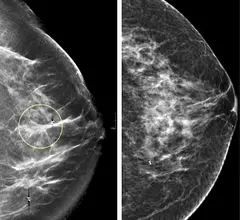
Comparison of a 2D digital mammogram and breast tomosynthesis 3D mammography from UCSF.

DBT exams take twice as long to read as 2D mammograms, but the learning curve is short

Although DBT exams are proven to identify more difficult to detect cancers, especially among women with dense breasts, they also include significantly more images than standard 2D mammograms. 

Thumbnail

New study results 'strongly' back supplemental MRI for women with dense breasts

This latest research further confirms that breast MRI not only detects tumors that mammography cannot, but it also spots invasive cases that pose greater risks to patients. 

partners.jpg

Bayer, Hologic unite to improve availability of contrast-enhanced mammography

The two companies have partnered on a new FDA-cleared CT injection system for the early detection of breast cancer.

medicare advantage rural healthcare hospitals

ACR chief research officer tapped for clinical trial recruitment initiative

Etta Pisano, MD, will identify rural hospitals and help them to onboard patients into new clinical studies.

Video of Samir Patel, MD, diagnostic radiologist at Radiology Inc., value management program founder and director and a board member of the Beacon Health System, explaining how AI is being implemented in mammography at RSNA 2023.

Real-world implementation of AI in mammography

Samir Patel, MD, radiologist at Radiology Inc., and a board member of the Beacon Health System, explains how AI is being implemented in clinical practice for breast imaging. 


Video interview with Radiology Business Management Association (RBMA) President Kit Crancer, and executive director of the Rayus Quality Institute, where he said the lack of federal policy on insurance coverage for diagnostic breast exams as opposed to screening exams

State legislation addresses gaps in breast imaging coverage, ensures affordable diagnostic exams

RBMA President Kit Crancer said a lack of federal policy has driven 20 states to adopt laws requiring that insurance cover the costs. 

Around the web

The nuclear imaging isotope shortage of molybdenum-99 may be over now that the sidelined reactor is restarting. ASNC's president says PET and new SPECT technologies helped cardiac imaging labs better weather the storm.

CMS has more than doubled the CCTA payment rate from $175 to $357.13. The move, expected to have a significant impact on the utilization of cardiac CT, received immediate praise from imaging specialists.

The all-in-one Omni Legend PET/CT scanner is now being manufactured in a new production facility in Waukesha, Wisconsin.