Breast Imaging

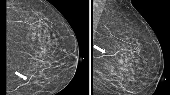
Breast imaging includes imaging modalities used for breast cancer screenings and planning therapy once cancer is detected. Mammography is the primary modality used. Mammogram technology is moving from 2D full-field digital mammography (FFDM) to breast tomosynthesis, or 3D mammography, which helps reduce false positive exams by allowing radiologists to look through the layers of tissue. Overlapping areas of dense breast tissue on 2D mammograms appear similar to cancers and 3D tomo helps determine if suspect areas are cancer or not. About 50% of women have dense breast tissue, which appears white on mammograms, the same as cancers, making diagnosis difficult. Radiologists use the Breast Imaging Reporting and Data System (BI-RADS) scoring system to define the density of breast tissue. Many states now require patients to be notified if they have dense breasts so they understand their mammograms might be suboptimal and they should use supplemental imaging that can see through the dense areas. This includes tomosythesis, breast ultrasound, automated breast ultrasound (ABUS), breast MRI, contrast enhanced mammography and nuclear imaging, including positron emission mammography (PEM).
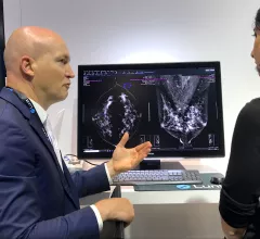
AI is still one of the key technologies on the floor many radiologists want to learn more about. of the AI. A product rep discussing breast automated detection AI in the crowded Lunit booth at RSNA 2022.

9 technology trends and takeaways from RSNA 2022

Here are some interesting new technologies and key trends from the vast expo floor at the Radiological Society of North America 2022 meeting.

Thumbnail

Google Health partners with iCAD in commercial AI imaging push

The deal is the first commercial partnership for Google Health to introduce its breast imaging AI into clinical practice.

Physician Senogram

Opto-acoustic Imaging: A new modality changing the future of breast imaging

Sponsored by Seno Medical

When it comes to breast imaging, a few new technologies have improved the visualization of lesions and diagnostic procedures. But a new modality could change the game completely.

Thumbnail

Can standard radiation treatments replace surgery for some breast cancer patients?

As chemotherapy treatments improve, patients who respond well may be able to skip surgery with a low chance of recurrence.

Constance "Connie" Lehman, MD, PhD,, chief of breast imaging, co-director of the Avon Comprehensive Breast Evaluation Center at the Massachusetts General Hospital, and professor of radiology at Harvard Medical School, discusses trends in breast imaging.

VIDEO: Connie Lehman discusses trends in breast imaging

Connie Lehman, MD, PhD, chief of breast imaging, co-director of the Avon Comprehensive Breast Evaluation Center at the Massachusetts General Hospital, and professor of radiology at Harvard Medical School, discusses trends she sees in breast imaging.

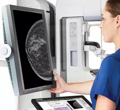
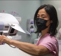
Breast screening mammogram during the COVID pandemic. COVID significantly impacted breast imaging. Image courtesy of Novant Health

VIDEO: Impact of COVID on breast imaging

Connie Lehman, MD, PhD, chief of breast imaging, co-director of the Avon Comprehensive Breast Evaluation Center at the Massachusetts General Hospital, discusses the challenges of the COVID-19 pandemic on breast screenings, increased cancer rates and issues with the vaccines causing false positives on mammograms.

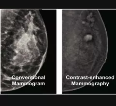
Contrast-enhanced mammography uses iodine contrast injected into a patient and mammography system to image contrast uptake or areas of increased vascular activity, which is typical of cancers. This can help image through dense breast tissue to find cancers that are otherwise masked by dense breast tissue.

VIDEO: Why contrast enhance mammography might be the ideal supplemental imaging

Connie Lehman, MD, PhD, chief of breast imaging, co-director of the Avon Comprehensive Breast Evaluation Center at the Massachusetts General Hospital, explains why she thinks contrast-enhanced mammography will likely become the go-to supplemental imaging modality for women with dense breasts.

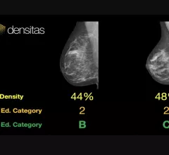
An example of commercially available artificial intelligence (AI) automated grading of breast density on mammograms from the vendor Densitas..

VIDEO: Role of AI in breast imaging with radiomics, detection of breast density and lesions

Connie Lehman, MD, chief of breast imaging, co-director of the Avon Comprehensive Breast Evaluation Center at Massachusetts General Hospital, discusses how artificial intelligence (AI) is being implemented in breast imaging.

Around the web

The ACR hopes these changes, including the addition of diagnostic performance feedback, will help reduce the number of patients with incidental nodules lost to follow-up each year.

And it can do so with almost 100% accuracy as a first reader, according to a new large-scale analysis.

The patient, who was being cared for in the ICU, was not accompanied or monitored by nursing staff during his exam, despite being sedated.