Magnetic Resonance Imaging

Magnetic resonance imaging (MRI) is the gold standard imaging modality for soft tissues. It produces detail cross-sectional images of soft tissue and bone anatomy, including muscles, tendons, ligaments, brain and organs, without the use of ionizing radiation. In addition to orthopedic imaging, MRI is also used for heart, brain and breast. MRI uses gadolinium contrast in many exams to highlight tissues and blood vessels, which enhances images and offers better diagnostic quality. It can also be used in conjunction with PET scans. How does MRI work? MR creates images by using powerful magnets to polarize hydrogen atoms in water (the body is made of of more than 80% water) so they face in one direction. A radiofrequency pulse is then used to ping these atoms, causing them to wobble, or resonate. The MRI coils detect this and computers can assemble images from the signals. Basic MRI scans will focus on the resonance of fat and water in two different sequences, which highlight and contrast different features in the anatomy.

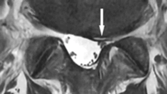
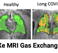
Sean Fain, PhD, vice chair of radiology and research and a professor of radiology, Division of Cardiovascular and Thoracic Imaging, University of Iowa, discusses how long-COVID lung damage can be tracked using xenon (Xe) gas magnetic resonance imaging (MRI) and quantitative computed tomography (CT). He spoke to Health Imaging at RSNA 2022.

VIDEO: Tracking long-COVID lung damage using MRI and CT

Sean Fain, PhD, vice chair of radiology and research and a professor of radiology, University of Iowa, discusses how long-COVID lung damage can be tracked using xenon (Xe) gas MRI and quantitative CT at RSNA 2022. 

Imaging industry names in the news: Koning, Medality, QT Imaging, Rezolut, Scanslated, more

Noteworthy market developments listed in the order announcements were posted.    

Why are women at lower risk for brain inflammation? Subcutaneous fat may hold the key

Researchers examined male and female mice on high-fat diets to learn about the impacts of fat distribution on brain inflammation. 

prostate cancer PSA

On its own, MRI-targeted biopsy takes big bite out of prostate cancer overdiagnosis

The gain came with the delayed discovery of only a few clinically significant cancers.

Cancer patients unruffled by whole-body MRI per se

Claustrophobic or not, most would choose the radiation-free modality over CT and tend to consider imaging-exam outcomes more worrisome than MRI in and of itself anyway.

Philips MRI

Growing contrast concerns accompany rising MRI volumes

More than half of surveyed radiologists worry about MRI contrast availability, yet almost all—99%—wish for contrast agents that would cut current gadolinium concentrations at least in half.

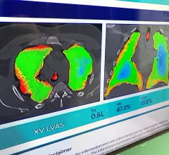
Dynamic lung air flow analysis just using X-ray without any contrast with new technology from 4D Medical.

PHOTO GALLERY: New technology and trends at RSNA 2022

Images from the Radiological Society of North America (RSNA) 2022 annual meeting Nov. 27- Dec. 1 in Chicago. The gallery includes new technologies and a look at sights around the world's largest radiology conference. 

kid child pediatric MRI imaging

Imaging research links obesity to poor brain health in children

The team will share its findings, based on imaging data from more than 5,000 pediatric patients, at RSNA 2022 in Chicago. 

Around the web

The nuclear imaging isotope shortage of molybdenum-99 may be over now that the sidelined reactor is restarting. ASNC's president says PET and new SPECT technologies helped cardiac imaging labs better weather the storm.

CMS has more than doubled the CCTA payment rate from $175 to $357.13. The move, expected to have a significant impact on the utilization of cardiac CT, received immediate praise from imaging specialists.

The all-in-one Omni Legend PET/CT scanner is now being manufactured in a new production facility in Waukesha, Wisconsin.