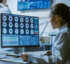
Magnetic Resonance Imaging

Magnetic resonance imaging (MRI) is the gold standard imaging modality for soft tissues. It produces detail cross-sectional images of soft tissue and bone anatomy, including muscles, tendons, ligaments, brain and organs, without the use of ionizing radiation. In addition to orthopedic imaging, MRI is also used for heart, brain and breast. MRI uses gadolinium contrast in many exams to highlight tissues and blood vessels, which enhances images and offers better diagnostic quality. It can also be used in conjunction with PET scans. How does MRI work? MR creates images by using powerful magnets to polarize hydrogen atoms in water (the body is made of of more than 80% water) so they face in one direction. A radiofrequency pulse is then used to ping these atoms, causing them to wobble, or resonate. The MRI coils detect this and computers can assemble images from the signals. Basic MRI scans will focus on the resonance of fat and water in two different sequences, which highlight and contrast different features in the anatomy.

Iodine contrast being loaded into a contrast injector in preparation for a cardiac CT scan at Duly Health and Care in Lisle, Illinois. The contrast shortage is causing some healthcare organizations to postpone exams and procedures and ration contrast supplies. Photo by Dave Fornell

‘Relatively new’ contrast vendor releases second generic agent

A global tech and pharma supplier that introduced a generic equivalent to GE Healthcare’s Visipaque during the height of the 2022 contrast shortage has begun offering a substitute for Guerbet’s Dotarem.

Female Medical Research Scientist Working with Brain Scans

FDA approves AI analysis of high-grade gliomas

An AI startup in the neuro-oncology space has received the government’s go-ahead to market software for analyzing certain fast-growing brain tumors on MRI.

Thumbnail

FDA clears 2D image-reconstruction product for 3D MRI

GE Healthcare may ship its deep learning-based image reconstruction software called AIR Recon DL with new 3D capabilities, the company announced Sept. 29.

Thumbnail

New amyloid buster significantly hinders Alzheimer’s advance

An experimental Alzheimer’s drug therapy has slowed cognitive and functional decline by 27% versus placebo in a double-blind, randomized study of 1,795 individuals with early signs and symptoms of the disease.

Thumbnail

For monitoring purposes, AI-aided MRI does what liver biopsy does with less risk, lower cost

Patients with autoimmune hepatitis may be better monitored across disease stages by AI-augmented multiparametric MRI than by liver biopsy, as the imaging has proven less costly and is inherently less risky due to its noninvasiveness. 

Could a repurposed drug break a centurylong stalemate in the war on cancer?

A pharmaceutical compound approved 21 years ago to treat breast cancer in postmenopausal women is showing fresh promise as a therapy for glioblastomas. 

Newborn brains invigorated by mother’s active lifestyle during pregnancy

The boon comes in the form of greater brain cortical thickness observable on neonatal MRI two weeks after the baby enters the world.

New contrast agent receives FDA approval after competitors collaborate on development

The Sept. 21 announcement states that Gadopiclenol Injection—a highly stable macrocyclic gadolinium-based contrast agent—is approved for use in adult and pediatric patients aged 2 years and older during MRI exams.

Around the web

The nuclear imaging isotope shortage of molybdenum-99 may be over now that the sidelined reactor is restarting. ASNC's president says PET and new SPECT technologies helped cardiac imaging labs better weather the storm.

CMS has more than doubled the CCTA payment rate from $175 to $357.13. The move, expected to have a significant impact on the utilization of cardiac CT, received immediate praise from imaging specialists.

The all-in-one Omni Legend PET/CT scanner is now being manufactured in a new production facility in Waukesha, Wisconsin.

Trimed Popup
Trimed Popup