Magnetic Resonance Imaging

Magnetic resonance imaging (MRI) is the gold standard imaging modality for soft tissues. It produces detail cross-sectional images of soft tissue and bone anatomy, including muscles, tendons, ligaments, brain and organs, without the use of ionizing radiation. In addition to orthopedic imaging, MRI is also used for heart, brain and breast. MRI uses gadolinium contrast in many exams to highlight tissues and blood vessels, which enhances images and offers better diagnostic quality. It can also be used in conjunction with PET scans. How does MRI work? MR creates images by using powerful magnets to polarize hydrogen atoms in water (the body is made of of more than 80% water) so they face in one direction. A radiofrequency pulse is then used to ping these atoms, causing them to wobble, or resonate. The MRI coils detect this and computers can assemble images from the signals. Basic MRI scans will focus on the resonance of fat and water in two different sequences, which highlight and contrast different features in the anatomy.
Iodine contrast being loaded into a contrast injector in preparation for a cardiac CT scan at Duly Health and Care in Lisle, Illinois. The contrast shortage is causing some healthcare organizations to postpone exams and procedures and ration contrast supplies. Photo by Dave Fornell

A new way to prepare imaging agents, thanks to a new method of radiolabeling

Isotopic labeling is time-consuming and expensive. Now, a new development in isotopic labeling heralds a new way to prepare imaging agents. 

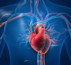
By visualizing metabolism, researchers hope to improve heart disease diagnosis

Hyperpolarized MRI may help identify metabolism problems—which can be an early signal of a heart condition—in real time.

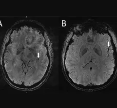
Ultra-high-res MRI Reveals Migraine Brain Changes

7T MRI scans provide a high-res look at how migraines affect the brain

In a new analysis, scheduled to be presented at RSNA 2022 in Chicago, researchers have identified enlarged perivascular spaces in the brains of migraine patients. This may be suggestive of waste clearance issues in the central nervous system.

Thumbnail

Experts ID imaging biomarkers in kids with ADHD

The study's authors explained how MRI results can be used to help identify children with ADHD—and how these findings could be used in the AI algorithms of tomorrow. The team will be presenting its findings at RSNA 2022 in Chicago. 

Canadian radiologists push federal lawmakers to deal with ‘devastating impact’ of long waits for medical imaging

Radiologists from across Canada descended on Parliament Hill in Ottawa earlier this month to voice pressing concerns on behalf of medical-imaging patients and professionals.

Lyme disease neuroimaging uncovers compensatory brain repair

Lyme disease patients treated for “brain fog” may develop compensatory alterations in white matter that show up on MRI and correspond—unexpectedly—with slow but sound cognitive performance.

Thumbnail

Neuroimaging recedes behind other cost compilers in stroke care

Contrary to older research that showed neuroimaging emerging as the single most dominating cost contributor in ischemic stroke care for older Americans, a new study shows treatment and other line items account for bigger slices of the bill.

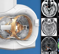
MRI system advances include helium-free MRI systems (left is Philips version of this system), and compressed sensing (right, GE's version of this technology), which can greatly reduce scan times. Both technologies were mentioned in an overview by Signet Research of new tech that will be at RSNA 2022.

VIDEO: Overview of MRI market and technology trends

Bhvita Jani, research manager, Signify Research, explains some key trends and technology advances in the magnetic resonance imaging (MRI) market leading into the Radiological Society of North America (RSNA) 2022 meeting. 

Around the web

The nuclear imaging isotope shortage of molybdenum-99 may be over now that the sidelined reactor is restarting. ASNC's president says PET and new SPECT technologies helped cardiac imaging labs better weather the storm.

CMS has more than doubled the CCTA payment rate from $175 to $357.13. The move, expected to have a significant impact on the utilization of cardiac CT, received immediate praise from imaging specialists.

The all-in-one Omni Legend PET/CT scanner is now being manufactured in a new production facility in Waukesha, Wisconsin.

Trimed Popup
Trimed Popup