Magnetic Resonance Imaging

Magnetic resonance imaging (MRI) is the gold standard imaging modality for soft tissues. It produces detail cross-sectional images of soft tissue and bone anatomy, including muscles, tendons, ligaments, brain and organs, without the use of ionizing radiation. In addition to orthopedic imaging, MRI is also used for heart, brain and breast. MRI uses gadolinium contrast in many exams to highlight tissues and blood vessels, which enhances images and offers better diagnostic quality. It can also be used in conjunction with PET scans. How does MRI work? MR creates images by using powerful magnets to polarize hydrogen atoms in water (the body is made of of more than 80% water) so they face in one direction. A radiofrequency pulse is then used to ping these atoms, causing them to wobble, or resonate. The MRI coils detect this and computers can assemble images from the signals. Basic MRI scans will focus on the resonance of fat and water in two different sequences, which highlight and contrast different features in the anatomy.

Tough sledding for patients using hospital ‘transparency’ tools to obtain imaging price estimates

Notwithstanding CMS’ most-shoppable-services rule, radiology researchers find much work lies ahead “if the goal of online price estimators is for patients to use them.”

Long and longer: Imaging wait times in Canada

Prior to the arrival of COVID-19 in early 2020, Canadian patients waited nearly three months for an MRI and more than 11 weeks for a CT. Things have only gotten worse since then.

‘Low-trust’ providers inadvertently increase pain in patients undergoing diagnostic testing

The effect shows up on functional MRI as increased brain activity in regions involved in pain, emotion and attention—not only during the procedure but also afterward, when patients remember the experience and score its discomfort level.

Thumbnail

5 imaging modalities have changed the war on cancer over the past 50 years. How many can you name?

Two branch chiefs with the cancer imaging program at NIH’s National Cancer Institute have produced a written commemoration to mark the golden anniversary of 1971’s National Cancer Act.

Emergency overreads change care for 92% of patients with discrepant outside reports

When emergency radiologists overread interpretations of critically ill transfer patients, they discover discrepancies in fewer than 15% of cases. However, more than 90% of these second opinions produce a change in patient care or follow-up.

FDA greenlights ortho robot, automated MRI brain modeling

FDA approvals arrive for ClearPoint Neuro and MicroPort Navibot. 

Thumbnail

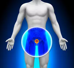
Prostate AI cleared for U.S. sales

A medical AI startup in Omaha, Neb., has received the FDA’s blessing to market software for diagnosing prostate cancer on MRI scans.

Around the web

The nuclear imaging isotope shortage of molybdenum-99 may be over now that the sidelined reactor is restarting. ASNC's president says PET and new SPECT technologies helped cardiac imaging labs better weather the storm.

CMS has more than doubled the CCTA payment rate from $175 to $357.13. The move, expected to have a significant impact on the utilization of cardiac CT, received immediate praise from imaging specialists.

The all-in-one Omni Legend PET/CT scanner is now being manufactured in a new production facility in Waukesha, Wisconsin.

Trimed Popup
Trimed Popup