Magnetic Resonance Imaging

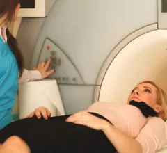
Magnetic resonance imaging (MRI) is the gold standard imaging modality for soft tissues. It produces detail cross-sectional images of soft tissue and bone anatomy, including muscles, tendons, ligaments, brain and organs, without the use of ionizing radiation. In addition to orthopedic imaging, MRI is also used for heart, brain and breast. MRI uses gadolinium contrast in many exams to highlight tissues and blood vessels, which enhances images and offers better diagnostic quality. It can also be used in conjunction with PET scans. How does MRI work? MR creates images by using powerful magnets to polarize hydrogen atoms in water (the body is made of of more than 80% water) so they face in one direction. A radiofrequency pulse is then used to ping these atoms, causing them to wobble, or resonate. The MRI coils detect this and computers can assemble images from the signals. Basic MRI scans will focus on the resonance of fat and water in two different sequences, which highlight and contrast different features in the anatomy.

AI startup competition tackles MRI times, cell imaging technology

Tech company Nvidia’s Inception contest seeks to find the top artificial intelligence (AI) startups in key sectors, including healthcare, to alter the direction of select industries.

fMRI shows 3-year-olds have brain networks to understand others

A team of researchers at the Massachusetts Institute of Technology (MIT) used functional MRI (fMRI) to explore how young children develop an ability known as “theory of mind,” which allows one person to infer about another’s mental state.

Thumbnail

Researchers find quick fix for RV parameters in pediatric CMR imaging

A group of doctors in Warsaw, Poland, have identified a single parameter for first-line assessment of right ventricular (RV) function in pediatric patients, according to data published in Clinical Radiology this week.

Thumbnail

Why do people miss MRIs? Race, money might have something to do with it

The greater the window of time between scheduling an MRI and showing up for the exam, the more likely a patient is to miss the appointment altogether, according to data published in the Journal of the American College of Cardiology—and the effect is especially pronounced in minority populations.

Thumbnail

3T MRI can alter hearing ability for weeks after exam

Less than an hour of 3T MRI exposure could result in weeks of altered hearing ability, even if a patient wears hearing protection during the exam, researchers reported in Radiology this month.

Philips debuts new MR system at ECR 2018

Royal Philips debuted its newest MR solution, the Ingenia Elition, on Wednesday, Feb. 28, at the European Congress of Radiology (ECR) 2018 in Vienna.

Thumbnail

Follow-up MRI imaging recommended for breast biopsies canceled due to nonvisualization

When MRI-guided breast biopsy is canceled due to nonvisualization, follow-up imaging typically finds that the lesion has completely resolved, according to a new study published in Academic Radiology. The authors still recommend follow-up MRI imaging six months later, though, because some lesions do persist.

Radiology practice suing MRI provider for $68K in late payments

American Radiology Technologies (ART), a Pinecrest, Florida-based radiology practice, is suing Extremity Imaging Partners (EIP) and Extremity MRI in Wexford, Pennsylvania, for allegedly failing to pay for services.

Around the web

The nuclear imaging isotope shortage of molybdenum-99 may be over now that the sidelined reactor is restarting. ASNC's president says PET and new SPECT technologies helped cardiac imaging labs better weather the storm.

CMS has more than doubled the CCTA payment rate from $175 to $357.13. The move, expected to have a significant impact on the utilization of cardiac CT, received immediate praise from imaging specialists.

The all-in-one Omni Legend PET/CT scanner is now being manufactured in a new production facility in Waukesha, Wisconsin.