Magnetic Resonance Imaging

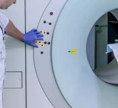
Magnetic resonance imaging (MRI) is the gold standard imaging modality for soft tissues. It produces detail cross-sectional images of soft tissue and bone anatomy, including muscles, tendons, ligaments, brain and organs, without the use of ionizing radiation. In addition to orthopedic imaging, MRI is also used for heart, brain and breast. MRI uses gadolinium contrast in many exams to highlight tissues and blood vessels, which enhances images and offers better diagnostic quality. It can also be used in conjunction with PET scans. How does MRI work? MR creates images by using powerful magnets to polarize hydrogen atoms in water (the body is made of of more than 80% water) so they face in one direction. A radiofrequency pulse is then used to ping these atoms, causing them to wobble, or resonate. The MRI coils detect this and computers can assemble images from the signals. Basic MRI scans will focus on the resonance of fat and water in two different sequences, which highlight and contrast different features in the anatomy.

Thumbnail

Machine learning, fMRI offer insight into OCD patients’ response to therapy

A team of California researchers has developed a method for predicting the responses of obsessive compulsive disorder (OCD) patients to cognitive behavioral therapy using machine learning and fMRI, according to work published in the journal PNAS.

Thumbnail

Mid-chemo MRI could predict early response to treatment in rectal cancer patients

Additional MR imaging performed during chemoradiation therapy could be an early predictor of a rectal cancer patient’s pathological response to treatment, according to research out of San Raffaele Hospital in Milan.

Thumbnail

Expansive data set of brain MRIs could guide treatment, recovery in stroke patients

One of the largest open-source data sets of brain MRIs from stroke patients is now available for public download via Scientific Data, a team of University of Southern California scientists reported this week.

FDA clears 3D MRI application from Siemens

Siemens announced Monday, Feb. 19, that the FDA has cleared the company’s GOKnee3D MRI application.

Imaging technique shows success as alternative to MRI in restless children

Scientists have found a viable alternative to conventional MR imaging in pediatric patients who have a hard time keeping still during exams, according to a study published this month in the American Journal of Roentgenology.

Thumbnail

Breast MRI is uncommon when treating male patients, but can still provide value

Radiologists rarely turn to breast MRI when treating male patients, but according to a new study published in Current Problems in Diagnostic Radiology, it can be effective in certain scenarios.

Thumbnail

‘Silent’ MRIs making progress in clinical settings; MRAs lag behind

Silent alternatives to conventional MRI scans are showing promising results, but reducing noise in MR angiograms (MRAs) is proving a more difficult task, a team of Stanford University scientists reported in the American Journal of Roentgenology this week.

Update: Imaging society says doctors should not be punished for MRI-related death

A branch of the Indian Radiological and Imaging Association (IRIA) has penned a letter defending the physicians arrested for their role in the MRI-related death of 32-year-old Rajesh Maruti Maru in Mumbai, India.

Around the web

The nuclear imaging isotope shortage of molybdenum-99 may be over now that the sidelined reactor is restarting. ASNC's president says PET and new SPECT technologies helped cardiac imaging labs better weather the storm.

CMS has more than doubled the CCTA payment rate from $175 to $357.13. The move, expected to have a significant impact on the utilization of cardiac CT, received immediate praise from imaging specialists.

The all-in-one Omni Legend PET/CT scanner is now being manufactured in a new production facility in Waukesha, Wisconsin.