Magnetic Resonance Imaging

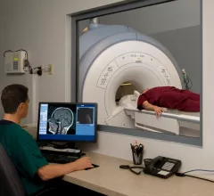
Magnetic resonance imaging (MRI) is the gold standard imaging modality for soft tissues. It produces detail cross-sectional images of soft tissue and bone anatomy, including muscles, tendons, ligaments, brain and organs, without the use of ionizing radiation. In addition to orthopedic imaging, MRI is also used for heart, brain and breast. MRI uses gadolinium contrast in many exams to highlight tissues and blood vessels, which enhances images and offers better diagnostic quality. It can also be used in conjunction with PET scans. How does MRI work? MR creates images by using powerful magnets to polarize hydrogen atoms in water (the body is made of of more than 80% water) so they face in one direction. A radiofrequency pulse is then used to ping these atoms, causing them to wobble, or resonate. The MRI coils detect this and computers can assemble images from the signals. Basic MRI scans will focus on the resonance of fat and water in two different sequences, which highlight and contrast different features in the anatomy.

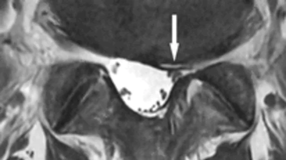
Breast MRI screening associated with higher biopsy rates, lower cancer yield findings than mammography alone

Patients with and without a personal history of breast cancer (PHBC) have higher biopsy rates and lower cancer yields following biopsy in the 90 days after screening MRI than in the 90 days after a traditional mammogram, according to a new study published in JAMA Internal Medicine.

Patient preparing to sue hospital after experiencing MRI-related burn in 2016

A 72-year-old patient from Glendale, California, is preparing to sue Glendale Adventist Medical Center for a burn he experienced before getting an MRI back in 2016.

Update: Investigation into MRI-related death hits a roadblock

The investigation into the MRI-related death of 32-year-old Rajesh Maruti Maru in Mumbai, India, continues, but police say they lack video evidence of the incident.

Sticker shock: Army vet gets $8K bill for MRI

As healthcare costs outpace inflation, patients are more and more cost-conscious—with many often shopping around to minimize out-of-pocket costs—but medicine remains far from transparent. A recent story from WBRC in Birmingham, Alabama, explored one man’s shock when he received an $8,000 bill for two MRIs on his back.

Thumbnail

Pre-biopsy prostate MRI utilization is up, but researchers see room for improvement

The utilization of pre-biopsy prostate MRI is up significantly among Medicare beneficiaries, according to a new study published in The Journal of Urology. But should that percentage be even higher?

Update: Radiologist arrested in connection with MRI-related death

A radiologist has been arrested in connection with the MRI-related death of 32-year-old Rajesh Maruti Maru.

Thumbnail

How well can MRI detect lymph node metastasis in patients with bladder and prostate cancer?

MRI can detect lymph node (LN) metastasis in patients with bladder and prostate cancer with high specificity, but its sensitivity is “poor and heterogeneous,” according to systematic review published in the American Journal of Roentgenology. Using ultrasmall superparamagnetic particles of iron oxide (USPIO) is one way providers can improve sensitivity.

Fallout continues in India after MRI-related death

A man from Mumbai was sucked into an MRI machine and died Jan. 27. Now, both the equipment’s manufacturer and the city’s government are planning to investigate the incident.

Around the web

The nuclear imaging isotope shortage of molybdenum-99 may be over now that the sidelined reactor is restarting. ASNC's president says PET and new SPECT technologies helped cardiac imaging labs better weather the storm.

CMS has more than doubled the CCTA payment rate from $175 to $357.13. The move, expected to have a significant impact on the utilization of cardiac CT, received immediate praise from imaging specialists.

The all-in-one Omni Legend PET/CT scanner is now being manufactured in a new production facility in Waukesha, Wisconsin.