Magnetic Resonance Imaging

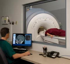
Magnetic resonance imaging (MRI) is the gold standard imaging modality for soft tissues. It produces detail cross-sectional images of soft tissue and bone anatomy, including muscles, tendons, ligaments, brain and organs, without the use of ionizing radiation. In addition to orthopedic imaging, MRI is also used for heart, brain and breast. MRI uses gadolinium contrast in many exams to highlight tissues and blood vessels, which enhances images and offers better diagnostic quality. It can also be used in conjunction with PET scans. How does MRI work? MR creates images by using powerful magnets to polarize hydrogen atoms in water (the body is made of of more than 80% water) so they face in one direction. A radiofrequency pulse is then used to ping these atoms, causing them to wobble, or resonate. The MRI coils detect this and computers can assemble images from the signals. Basic MRI scans will focus on the resonance of fat and water in two different sequences, which highlight and contrast different features in the anatomy.

Arrests made after man dies being sucked into MRI chamber

A 32-year-old man from Mumbai, India, died on Jan. 27 after being sucked into an MRI while holding a metallic oxygen cylinder in his hand. Three BYL Nair Municipal Hospital employees have been arrested.

Canon Medical Systems installs new MR system in Ohio research center

Canon Medical Systems USA announced today that, as part of its partnership with Quality Electrodynamics (QED), the company has installed a Vantage Galan 3T MR system at the new QED Research Center in Mayfield Village, Ohio.

MRIs found safe for patients with implantable cardiac devices

Patients fitted with implantable cardiac devices—both approved and unapproved by the FDA—should steer clear of outdated MRI safety information. A recent study released by the New England Journal of Medicine found these patients are safe to undergo MRI screenings, as reported in a recent article by Cardiovascular Business. 

Thumbnail

What impact does MRI, CT utilization have on ACOs?

CMS data from accountable care organizations (ACOs) participating in the Medicare Shared Savings Program (MSSP) reveals that higher MRI utilization rates are associated with “small but significant” cost savings, according to a recent analysis published in the Journal of the American College of Radiology. No such correlation was seen for CT utilization rates.

Patients prefer TVUS to MRI for pelvic imaging

Providers often turn to transvaginal ultrasound (TVUS) and MRI when women are experiencing pelvic pain or abnormal uterine bleeding. Which of these imaging modalities is preferred among women? A team of researchers studied that exact topic, publishing their findings in Academic Radiology.

Patient shares struggles with being claustrophobic and undergoing MRI

It’s a problem radiologists and technologists encounter from time to time: the claustrophobic patient who needs an MRI. They need the exam, but they’re not exactly happy about the process.

Thumbnail

RSNA 2017: Overuse of STAT designation slows MRI workflow, causes confusion

The term “STAT” implies urgency: act immediately. But when clinicians overuse that designation, it becomes more difficult and time-consuming to distinguish true emergencies from cases that can wait, said Anna Trofimova, MD, PhD, a diagnostic radiology resident at Emory University School of Medicine.

Toshiba Medical Launches New MR to Enhance Patient Comfort in Community Hospital Settings

CHICAGO, Nov. 28, 2017 – Addressing patients’ top MR imaging anxieties, Toshiba Medical, a Canon Group company, introduces its newest MR system, the Vantage Elan™ / Zen Edition 1.5T to deliver comfortable, effortless and efficient exams in healthcare facilities of all sizes. The Zen Edition prioritizes intelligent work­flow and patient comfort, providing clinical applications to help providers make accurate diagnoses.

Around the web

The nuclear imaging isotope shortage of molybdenum-99 may be over now that the sidelined reactor is restarting. ASNC's president says PET and new SPECT technologies helped cardiac imaging labs better weather the storm.

CMS has more than doubled the CCTA payment rate from $175 to $357.13. The move, expected to have a significant impact on the utilization of cardiac CT, received immediate praise from imaging specialists.

The all-in-one Omni Legend PET/CT scanner is now being manufactured in a new production facility in Waukesha, Wisconsin.