Magnetic Resonance Imaging

Magnetic resonance imaging (MRI) is the gold standard imaging modality for soft tissues. It produces detail cross-sectional images of soft tissue and bone anatomy, including muscles, tendons, ligaments, brain and organs, without the use of ionizing radiation. In addition to orthopedic imaging, MRI is also used for heart, brain and breast. MRI uses gadolinium contrast in many exams to highlight tissues and blood vessels, which enhances images and offers better diagnostic quality. It can also be used in conjunction with PET scans. How does MRI work? MR creates images by using powerful magnets to polarize hydrogen atoms in water (the body is made of of more than 80% water) so they face in one direction. A radiofrequency pulse is then used to ping these atoms, causing them to wobble, or resonate. The MRI coils detect this and computers can assemble images from the signals. Basic MRI scans will focus on the resonance of fat and water in two different sequences, which highlight and contrast different features in the anatomy.

Siemens Healthineers announces FDA clearance of 3T MRI System

Siemens Healthineers announced Thursday that the FDA has cleared the company’s MAGNETOM Vida 3 Tesla (3T) MRI scanner. The scanner features Siemens’ new BioMatrix technology, which “addresses inherent anatomical and physiological differences among patients,” and a user interface designed to simplify positioning the patient table.

Thumbnail

Breast MR in community practices meets ACR benchmarks

A study of over 13,000 MR breast screening exams determined the modality was meeting performance benchmarks set by the American College of Radiology, but found room for improvement on false-positive results, according to an article published online in Radiology.

MRI is a low-dose option for diagnosing Crohn's study finds

MRI could replace CT as a low-dose option when diagnosing Crohn’s Disease in the small bowel, according to a study published in Academic Radiology.

MRI before biopsy may be cost-effective detection method for prostate cancer

Researchers from Case Western Reserve University School of Medicine and University Hospitals Cleveland Medical Center revealed in a new study that diagnostic MRI followed by one of three MRI-guided biopsy strategies could be a cost-effective detection method for prostate cancer.

MRI study links weight loss, slowed degeneration of knee cartilage

Researchers, using MRI on overweight and obese patients, found that individuals who lost a significant amount of weight over a four-year period showed significantly less degeneration of their knee cartilage.

Toshiba Medical’s Vantage Galan 3T MR Delivers Fast, Accurate Results

Offering a full range of advanced clinical applications for researchers, Toshiba Medical will demonstrate its most advanced MR technology, the Vantage GalanTM 3T, at this year’s International Society for Magnetic Resonance in Medicine (ISMRM) 2017 annual meeting in Honolulu, Hawaii, April 22–27, 2017 (Booth #401).

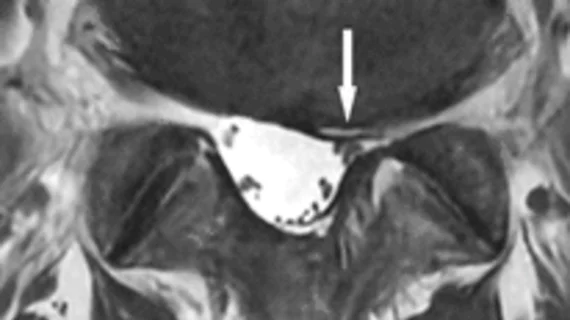
Variation in spinal MRI can muddle employer-based health plans

Costs of imaging procedures are often compared to one another when medical price shopping. It’s instinctive: All MRI machines are more or less the same, right? However, a study published in Spine Journal found a tenfold difference in price among 10 different imaging centers across the country.

fMRI shows beetroot juice before exercise helps older brains

Using resting-state MRI functional brain network organization, scientists at Wake Forest University revealed that drinking a beetroot juice supplement before exercising improves performance in the brain of older individuals, mirroring the operations of a younger brain.

Around the web

The nuclear imaging isotope shortage of molybdenum-99 may be over now that the sidelined reactor is restarting. ASNC's president says PET and new SPECT technologies helped cardiac imaging labs better weather the storm.

CMS has more than doubled the CCTA payment rate from $175 to $357.13. The move, expected to have a significant impact on the utilization of cardiac CT, received immediate praise from imaging specialists.

The all-in-one Omni Legend PET/CT scanner is now being manufactured in a new production facility in Waukesha, Wisconsin.