Magnetic Resonance Imaging

Magnetic resonance imaging (MRI) is the gold standard imaging modality for soft tissues. It produces detail cross-sectional images of soft tissue and bone anatomy, including muscles, tendons, ligaments, brain and organs, without the use of ionizing radiation. In addition to orthopedic imaging, MRI is also used for heart, brain and breast. MRI uses gadolinium contrast in many exams to highlight tissues and blood vessels, which enhances images and offers better diagnostic quality. It can also be used in conjunction with PET scans. How does MRI work? MR creates images by using powerful magnets to polarize hydrogen atoms in water (the body is made of of more than 80% water) so they face in one direction. A radiofrequency pulse is then used to ping these atoms, causing them to wobble, or resonate. The MRI coils detect this and computers can assemble images from the signals. Basic MRI scans will focus on the resonance of fat and water in two different sequences, which highlight and contrast different features in the anatomy.

Thumbnail

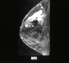
MRI useful for second look at inconclusive mammograms

MRI can be a useful tool for taking a second look at mammograms that are considered inconclusive in a process known as “problem-solving MRI," but radiologists should be cautious about the possibility of false negatives, according to a study published in the American Journal of Roentgenology.

Toshiba Medical’s Newest MRI System Tapped for UCI Brain Trauma Research

TUSTIN, Calif., March 8, 2017 – Researchers at the University of California, Irvine now have access to Toshiba Medical’s most advanced MR technology for their cutting edge neurology work investigating cerebral microbleeds (CMB) in high school football players as a potential precursor to chronic traumatic encephalopathy (CTE). As part of its ongoing, first-of-its-kind study to identify early signs of brain trauma in young athletes, UCI doctors will now leverage Toshiba Medical’s all-new Vantage GalanTM 3T MR system as a key diagnostic tool.

Siemens Healthineers Paves the Way for Precision Medicine With BioMatrix Technology and Magnetom Vida MRI Scanner

New BioMatrix technology adapts automatically to individual anatomical and physiological characteristics to offer consistent, high-quality imaging for all patients

Could a new contrast agent for MRI be on the horizon?

Massachusetts Institute of Technology (MIT) researchers developed a new specially coated iron oxide nanoparticle that could provide an alternative to conventional gadolinium-based contrast agents used for MRI procedures. Current gadolinium agents, have in rare cases, been found to create adverse effects in patients with impaired kidney function.

FDA Clears Cost-efficient MAGNETOM Sempra MRI System From Siemens Healthineers

1.5 Tesla, 60-cm MRI system offers innovative applications to improve competitiveness and potentially address profitability

MRI pioneer and Nobel prize winning physicist dies at 83

Peter Mansfield, physicist and winner of the Nobel Prize for his work in helping to invent MRI scanners died at the age of 83, according to a statement by the University of Nottingham.

Thumbnail

MRI prostate analysis could indicate cancer sans biopsy, reduce unnecessary procedures

An interdisciplinary team used an MRI quantification technique called "shape atlasing" to detect subtle deformations in cancerous prostate glands, finding a consistent anterior bulge among the patient cohort.

MRI illuminates changes in astronauts' gray matter

A new study has detailed what spaceflight can do to the shape and consistency of the human brain. Researchers examined structural MRIs of astronauts and found considerable changes in the gray matter in various sections of the brain.

Around the web

The nuclear imaging isotope shortage of molybdenum-99 may be over now that the sidelined reactor is restarting. ASNC's president says PET and new SPECT technologies helped cardiac imaging labs better weather the storm.

CMS has more than doubled the CCTA payment rate from $175 to $357.13. The move, expected to have a significant impact on the utilization of cardiac CT, received immediate praise from imaging specialists.

The all-in-one Omni Legend PET/CT scanner is now being manufactured in a new production facility in Waukesha, Wisconsin.