Magnetic Resonance Imaging

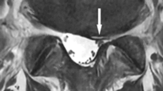
Magnetic resonance imaging (MRI) is the gold standard imaging modality for soft tissues. It produces detail cross-sectional images of soft tissue and bone anatomy, including muscles, tendons, ligaments, brain and organs, without the use of ionizing radiation. In addition to orthopedic imaging, MRI is also used for heart, brain and breast. MRI uses gadolinium contrast in many exams to highlight tissues and blood vessels, which enhances images and offers better diagnostic quality. It can also be used in conjunction with PET scans. How does MRI work? MR creates images by using powerful magnets to polarize hydrogen atoms in water (the body is made of of more than 80% water) so they face in one direction. A radiofrequency pulse is then used to ping these atoms, causing them to wobble, or resonate. The MRI coils detect this and computers can assemble images from the signals. Basic MRI scans will focus on the resonance of fat and water in two different sequences, which highlight and contrast different features in the anatomy.

Thumbnail

Breast MRI found to have a high sensitivity for pregnancy-associated breast cancer

Pregnancy-associated breast cancer (PABC) is rare, but aggressive. According to a recent study published in the American Journal of Roentgenology, breast MRI has a high sensitivity for PABC and changes the surgical management of PABC patients at a significant rate.

Thumbnail

MRI at center of Notre Dame lawsuit

Former Notre Dame linebacker Douglas Randolph is suing the university for allegedly concealing the results of an MRI originating from a hit he suffered during a practice in 2015.

Slow-progressing diseases detected earlier with MRI

MRI can be used to detect subtle changes in muscle tissue that allow early diagnosis of slowly progressing conditions, including facioscapulohumeral muscular dystrophy.

Thumbnail

MRI safety reminder: Cloth can be a fire hazard

Using cloth containing copper fibers during an MRI can result in a fire, as explained in a recent case report published by Radiology. The authors wrote their analysis about a specific incident that took place in August 2016 on a PET/MR imaging system that had just been installed the following year. The patient, a 61-year-old woman, was not harmed.

Thumbnail

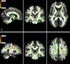
MRI connects genetic autism, structural abnormalities in brain

The duplication or deletion of a small piece of chromosome is one of the most common causes of genetic autism spectrum disorder. Researchers used MRI to examine 331 individuals to identify structural abnormalities in such brains.

Using breast MRI to evaluate newly diagnosed breast cancers does not delay treatment

Breast MRI is often used to evaluate newly diagnosed breast cancers, but there is some evidence that suggests this practice can lead to delayed surgical treatment. To explore that issue more closely, the authors of a recent study in the Journal of the American College of Radiology carried out a retrospective study of 189 patients who were diagnosed and treated for breast cancer at the same facility.

MRI offers less invasive peek at potential liver donors

Transplantation is the most effective method of combatting end-stage liver disease. But the success has increased demand and limited supplies for cadaveric organs. In a study, published online July 27 in the American Journal of Roentgenology, researchers examined how MRI can improve evaluation procedures of living liver donors.

Thumbnail

AI ‘learns’ to predict schizophrenia from brain MRI

A collaborative effort between IBM and the University of Alberta in Canada has produced artificial intelligence (AI) and machine learning algorithms were able to examine MRI and predict schizophrenia with 74 percent accuracy.

Around the web

The nuclear imaging isotope shortage of molybdenum-99 may be over now that the sidelined reactor is restarting. ASNC's president says PET and new SPECT technologies helped cardiac imaging labs better weather the storm.

CMS has more than doubled the CCTA payment rate from $175 to $357.13. The move, expected to have a significant impact on the utilization of cardiac CT, received immediate praise from imaging specialists.

The all-in-one Omni Legend PET/CT scanner is now being manufactured in a new production facility in Waukesha, Wisconsin.