Magnetic Resonance Imaging

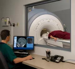
Magnetic resonance imaging (MRI) is the gold standard imaging modality for soft tissues. It produces detail cross-sectional images of soft tissue and bone anatomy, including muscles, tendons, ligaments, brain and organs, without the use of ionizing radiation. In addition to orthopedic imaging, MRI is also used for heart, brain and breast. MRI uses gadolinium contrast in many exams to highlight tissues and blood vessels, which enhances images and offers better diagnostic quality. It can also be used in conjunction with PET scans. How does MRI work? MR creates images by using powerful magnets to polarize hydrogen atoms in water (the body is made of of more than 80% water) so they face in one direction. A radiofrequency pulse is then used to ping these atoms, causing them to wobble, or resonate. The MRI coils detect this and computers can assemble images from the signals. Basic MRI scans will focus on the resonance of fat and water in two different sequences, which highlight and contrast different features in the anatomy.

ARRS to Host Abdominal and Pelvic MR Imaging Symposium in Baltimore

Leesburg, VA, June 15, 2016— ARRS, working in conjunction with its members, will hold the 2016 ARRS Abdominal and Pelvic MR Imaging Symposium Sept. 30–Oct. 1, 2016, at the Renaissance Baltimore Harborplace Hotel in downtown Baltimore. 

2016 ARRS Abdominal and Pelvic MR Imaging Symposium coming to Baltimore in September

The American Roentgen Ray Society (ARRS) previewed its 2016 ARRS Abdominal and Pelvic MR Imaging Symposium earlier this week. The symposium will be held at the Renaissance Baltimore Harborplace Hotel in Baltimore, Md., from Sept. 30 to Oct. 1, 2016.  

UCSF exhibit offers visitors a look at MRI history

The University of California, San Francisco (UCSF) library is now offering a sneak peek of its new Radiologic Imaging Laboratory (RIL) exhibit. 

Smart Choice MRI gains new $7 million investment

Smart Choice MRI, a Wisconsin-based healthcare provider known for providing MRIs for an all-inclusive fee of $600 or less, has received a $7 million investment from health system Edward-Elmhurst Health. 

Raymond V. Damadian, inventor of the MRI, to speak on May 17

Raymond V. Damadian, MD, inventor of the MRI, is scheduled to speak at the University of Buffalo in Buffalo, N.Y., on May 17. 

Thumbnail

Review tracks appropriateness—or lack thereof—of shoulder MRI orders

Shoulder MRI examinations are regularly not ordered according to existing appropriateness criteria, according to a new single-site study published by the Journal of the American College of Radiology.

Sharmila Majumdar honored with ISMRM 2016 Gold Medal

The International Society of Magnetic Resonance in Medicine (ISMRM) announced this week it has awarded its 2016 Gold Medal to Sharmila Majumdar, PhD, professor and vice chair for research of the department of radiology and biomedical imaging at the University of California, San Francisco (UCSF). Majumdar is also director of the Musculoskeletal and Quantitative Imaging (MQIR) group at UCSF. 

Radiologist’s new startup dramatically speeds up MR spectroscopy analysis

Thanks to a generous grant, a radiologist is now turning her years-old idea into a new healthcare startup. 

Around the web

The nuclear imaging isotope shortage of molybdenum-99 may be over now that the sidelined reactor is restarting. ASNC's president says PET and new SPECT technologies helped cardiac imaging labs better weather the storm.

CMS has more than doubled the CCTA payment rate from $175 to $357.13. The move, expected to have a significant impact on the utilization of cardiac CT, received immediate praise from imaging specialists.

The all-in-one Omni Legend PET/CT scanner is now being manufactured in a new production facility in Waukesha, Wisconsin.