Radiologists use diagnostic imaging to non-invasively look inside the body to help determine the causes of an injury or an illness, and confirm a diagnosis. Providers use many imaging modalities to do so, including CT, MRI, X-ray, Ultrasound, PET and more.
Chest CT is routinely performed for things like lung cancer screening, coronary calcium assessment and pulmonary nodule surveillance, but the modality also can be used to extract additional information.
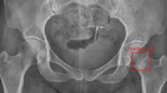
By highlighting subtle abnormalities that could otherwise be overlooked, an algorithm could potentially serve as an additional safety net when radiographic findings are indeterminate.
Shenzhen Second People's Hospital sits near the Wuhan area and has examined more than 1,400 suspected cases of COVID-19. Clinicians detailed their experience in Academic Radiology.
A new special report published in Radiology includes two new case studies of individuals infected with the illness, and details how experts can harness CT to help diagnose 2019-nCoV.
A team from Ho Chi Minh City in Vietnam recently detailed the case of a 65-year-old man who acquired the virus after traveling to Hanoi from the Wuchang district in Wuhan with his family.
Underfunding and a lack of resources have led to significant problems imaging severely injured patients in trauma centers across the UK's health system. The Royal College of Radiologists has called on the government to address the issues.
Patients who are enrolled in Ireland’s public health system are waiting, on average, 120 days more for a brain MRI compared to those who can afford private coverage, the Irish Times reports.