Radiologists use diagnostic imaging to non-invasively look inside the body to help determine the causes of an injury or an illness, and confirm a diagnosis. Providers use many imaging modalities to do so, including CT, MRI, X-ray, Ultrasound, PET and more.
Chest CT is routinely performed for things like lung cancer screening, coronary calcium assessment and pulmonary nodule surveillance, but the modality also can be used to extract additional information.
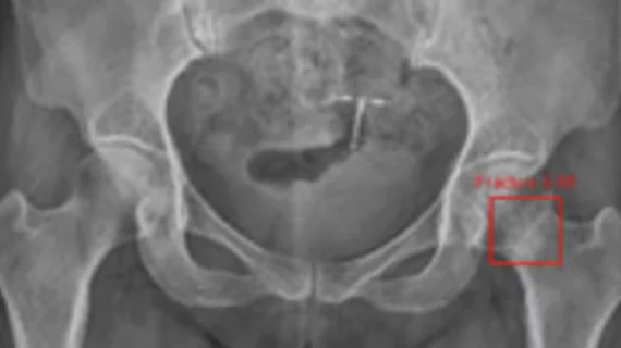
By highlighting subtle abnormalities that could otherwise be overlooked, an algorithm could potentially serve as an additional safety net when radiographic findings are indeterminate.
Not only can the metamaterial boost signal-to-noise ratio 10-fold, but It can reduce scan times, potentially making the modality more widely available for patients at lower costs.
Nearly one in five inductions of labor lead to a required emergency C-section, but current methods to predict such events are largely subjective with low predictive accuracy.
A new report published in Radiology: Cardiothoracic Imaging offers guidance for radiologists to help identify vaping product use-associated lung injury, analyzing recent cases and best practices.
Using the quiet protocol reduced noise levels and improved imaging success rates compared to conventional MRI, according to authors of a new study published in Academic Radiology.
The CDC released interim guidance for clinicians to help with the evaluation and management of e-cigarette or vaping product use-associated lung injury, or EVALI.
A new magnetic “seed” technology can help radiologists localize breast lesions and offers benefits over traditional wire-guided localization (WGL), reported authors of a study published Oct. 3 in the European Journal of Radiology.
MRI can help diagnose minor stroke and transient ischemic attacks (TIA) in patients who experience symptoms that aren’t always associated with stroke, according to a multi-year study published Sept. 23 in JAMA Neurology.