Radiologists use diagnostic imaging to non-invasively look inside the body to help determine the causes of an injury or an illness, and confirm a diagnosis. Providers use many imaging modalities to do so, including CT, MRI, X-ray, Ultrasound, PET and more.
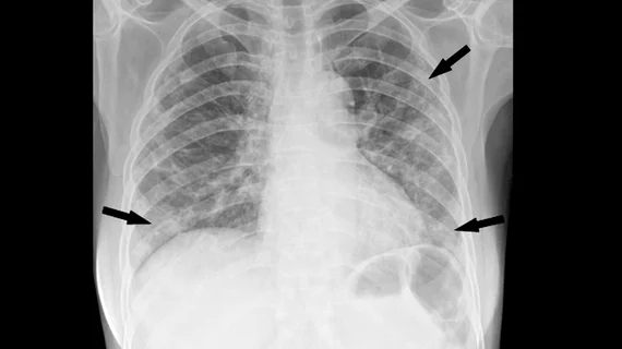
Two months after a cluster of cases occurred on a cruise ship, experts are offering insight into how radiologists can help spot the "silent" signs of the deadly virus on imaging.
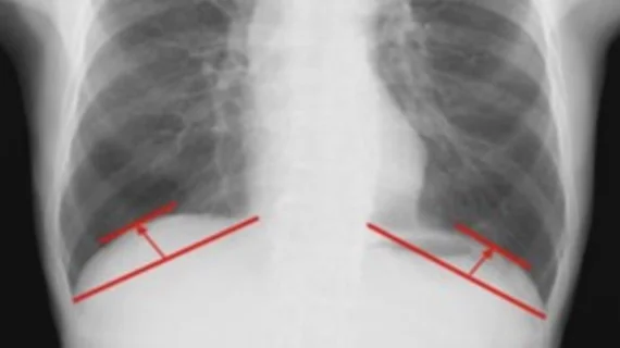
For the first time, research has identified diaphragmatic dome height measured on chest X-rays as an indicator of what patients’ post-op recovery will look like following surgery for cancer or other lung disorders.
On-the-field imaging helps determine if athletes can be treated effectively on-site or whether they might need to be transferred to a medical center for additional care.
The method—dual-energy CT virtual non-calcium (VNCa) imaging—can remove calcium from CT data and produce a quantitative assessment of injuries in the largest and most complex joint in the human body.
A deep neural network platform can help radiologists detect abdominal aortic aneurysms (AAAs) on CT images, and is especially helpful in clinically challenging cases, according to research presented at the SIIM annual conference.
A higher level of background parenchymal enhancement (BPE) measured during breast MRI is associated with the presence of breast cancer in women at high risk of breast cancer but not in women with average risk, according to a new study.
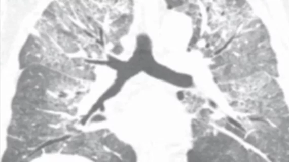
Enhanced dual-source dual-energy CT (DECT) can help differentiate invasive adenocarcinomas from preinvasive lesions which appear as pure ground-glass nodules (pGGNs), according to a small study published in the American Journal of Roentgenology.
“The results of this research are extremely exciting, as it will significantly impact clinical care,” reported study author Mishal Mendiratta-Lala, MD, with the division of abdominal radiology at Michigan Medicine in Ann Arbor.
The researchers analyzed the frequency and cancer yield of ACR Breast Imaging Reporting and Data System (BI-RADS) 3 lesions in patients who received baseline and non-baseline screening MRIs.
Some children in isolated villages have never seen an ultrasound machine, nevermind a portable one. A recent New York Times article provided an in-depth look at how impactful low-cost scanning technology can be to regions that don’t have access to basic imaging modalities.
High-strength 7T MRI can better track cortical brain lesions and play a crucial role in evaluating the progression of multiple sclerosis (MS), but some experts aren't sure it is clinically feasible.