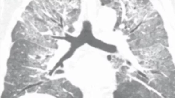
Radiologists use diagnostic imaging to non-invasively look inside the body to help determine the causes of an injury or an illness, and confirm a diagnosis. Providers use many imaging modalities to do so, including CT, MRI, X-ray, Ultrasound, PET and more.
Two months after a cluster of cases occurred on a cruise ship, experts are offering insight into how radiologists can help spot the "silent" signs of the deadly virus on imaging.
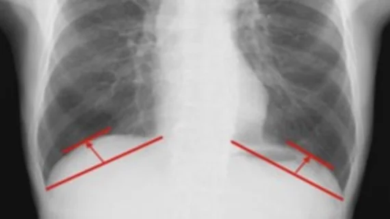
For the first time, research has identified diaphragmatic dome height measured on chest X-rays as an indicator of what patients’ post-op recovery will look like following surgery for cancer or other lung disorders.
On-the-field imaging helps determine if athletes can be treated effectively on-site or whether they might need to be transferred to a medical center for additional care.
The 40 evidence-based recommendations touch on everything from patient selection to study choice, radiation safety, clinical team dynamics, procedural techniques, study interpretations and more.
In a sampling of 2,273 chest radiographs of kids aged 2 to 18-years-old the AI-based software achieved diagnostic accuracies ranging from 86% to 96.9% for detecting a myriad of pathologies.
“These findings emphasize the importance of early recognition of IPV and timely intervention to prevent further harm to the victim,” authors of research published in Academic Radiology cautioned.
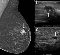
“The findings indicate that the DBT spot compression view can be performed in routine clinical practice to improve characterization of subtle or ambiguous findings on DBT,” experts wrote in AJR.
The $3.8 million grant spans five years and will focus on cerebrovascular abnormalities by using a specialized imaging technique, the university announced this week.