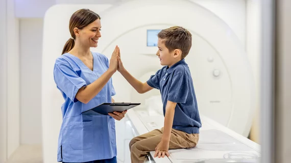
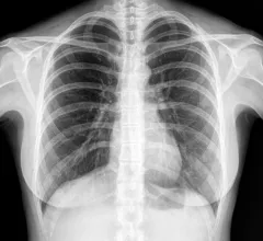
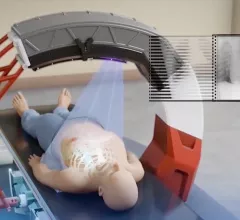
Radiologists use diagnostic imaging to non-invasively look inside the body to help determine the causes of an injury or an illness, and confirm a diagnosis. Providers use many imaging modalities to do so, including CT, MRI, X-ray, Ultrasound, PET and more.
Chest CT is routinely performed for things like lung cancer screening, coronary calcium assessment and pulmonary nodule surveillance, but the modality also can be used to extract additional information.
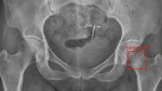
By highlighting subtle abnormalities that could otherwise be overlooked, an algorithm could potentially serve as an additional safety net when radiographic findings are indeterminate.
Radiologists used an AI tool-building platform to create their model(s), which allows clinicians the opportunity to develop AI models without any prior training in data sciences or computer programming.
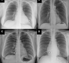
The tool’s sensitivity was recorded as 99.1% for abnormal radiographs and 99.8% for critical radiographs—better than two board-certified radiologists who also interpreted the exams.
Has point-of-care ultrasound outpaced hospitals’ capacity to incorporate the technology without anointing any particular specialty its proper guardian? The case could be made.
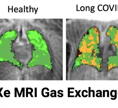
Sean Fain, PhD, vice chair of radiology and research and a professor of radiology, University of Iowa, discusses how long-COVID lung damage can be tracked using xenon (Xe) gas MRI and quantitative CT at RSNA 2022.
Tim Szczykutowicz, PhD, DABR, associate professor of radiology at the University of Wisconsin-Madison, is helping develop a new type of photon-counting CT detector that was shown as a work-in-progress by GE Healthcare at RSNA 2022.
The Israeli vendor Nanox says it has a vision for the future of healthcare. It seeks to address health disparities and access challenges with a new business model and innovative package of technologies. Hurdles loom, but opportunities abound.