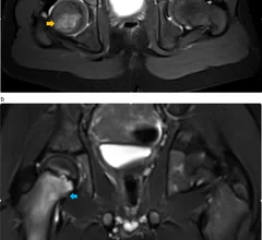
Radiologists use diagnostic imaging to non-invasively look inside the body to help determine the causes of an injury or an illness, and confirm a diagnosis. Providers use many imaging modalities to do so, including CT, MRI, X-ray, Ultrasound, PET and more.
Chest CT is routinely performed for things like lung cancer screening, coronary calcium assessment and pulmonary nodule surveillance, but the modality also can be used to extract additional information.
By highlighting subtle abnormalities that could otherwise be overlooked, an algorithm could potentially serve as an additional safety net when radiographic findings are indeterminate.
Serious discrepancies between preliminary imaging reads and final radiology reports are at risk of accumulating when the prelims are rendered during overnight hours.
Radiology education researchers have created an image-intensive online course for third- and fourth-year medical students wishing to learn radiology remotely.
Surveying the landscape of interpretive AI in radiology, two researchers note a yawning gap between great expectations set in the recent past and actual clinical implementations as of spring 2023.
Not only could the materials reduce patient exposure to ionizing radiation, they also could reduce costs associated with traditional X-ray equipment, according to newly published research in Nature Communications.
Experts from Mayo Clinic recently detailed their experience with the new offering, sharing that out of their 10 top ranked candidates, six had signaled the program.
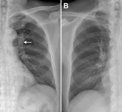
Features pertaining to location, density and superimposed structures were recently found to be associated with poorer outcomes for patients who initially had their lung cancer overlooked on radiographs.
The Australia-based company made the announcement on April 12 in a release that described the timing of these AI-assisted solutions as “increasingly important” amid growing workloads and staffing shortages.