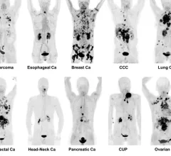
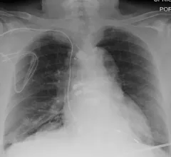
Radiologists use diagnostic imaging to non-invasively look inside the body to help determine the causes of an injury or an illness, and confirm a diagnosis. Providers use many imaging modalities to do so, including CT, MRI, X-ray, Ultrasound, PET and more.
Chest CT is routinely performed for things like lung cancer screening, coronary calcium assessment and pulmonary nodule surveillance, but the modality also can be used to extract additional information.
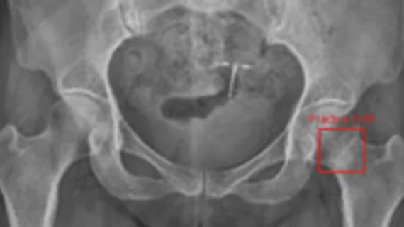
By highlighting subtle abnormalities that could otherwise be overlooked, an algorithm could potentially serve as an additional safety net when radiographic findings are indeterminate.
An artificial intelligence system that is currently commercially available for use in adults could also have applications in a pediatric population, according to a new study in Pediatric Radiology.
Munir Ghesani, MD, President of the Society of Nuclear Medicine and Molecular Imaging (SNMMI), system chief of nuclear medicine at Mount Sinai Health, explains recent advances in nuclear imaging technology.
After experts from one institution evaluated 500 portable chest x-rays completed during the summer of 2021, it was revealed that 46.2% of the images obtained were problematic, requiring the imaging to be repeated.
A new analysis offers a detailed comparison of soft-tissue lymphomas and soft-tissue tumors based on imaging characteristics from MRI scans—an area of study that has not yet been rigorously explored, the authors of the paper indicated.
When combined with artificial intelligence-based noise reduction techniques, new photon-counting CT technology can increase the detection of bone disease while also decreasing radiation exposure.
Authors of the new EJR paper explained that, although most of these tumors present in a similar way—with a lump or localized pain—their origins are wide-ranging and require the use of additional imaging to characterize the lesion.