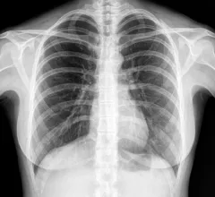
Radiologists use diagnostic imaging to non-invasively look inside the body to help determine the causes of an injury or an illness, and confirm a diagnosis. Providers use many imaging modalities to do so, including CT, MRI, X-ray, Ultrasound, PET and more.
Chest CT is routinely performed for things like lung cancer screening, coronary calcium assessment and pulmonary nodule surveillance, but the modality also can be used to extract additional information.
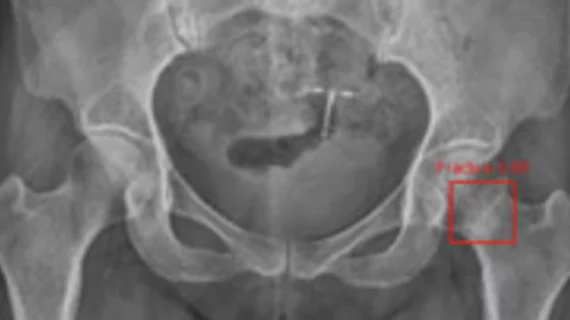
By highlighting subtle abnormalities that could otherwise be overlooked, an algorithm could potentially serve as an additional safety net when radiographic findings are indeterminate.
If left undiagnosed and untreated for a prolonged period, fatty liver disease can progress to more serious conditions, such as cirrhosis and liver cancer.
Following a recent surge of mycoplasma pneumoniae pneumonia cases, experts have issued new guidance to help providers quickly identify and treat the condition, with imaging playing a prominent role.
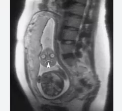
This is a clinical photo gallery of fetal imaging that explains what all can be seen on medical imaging, how sex is determined, how measurements are used to track the development of a baby.